The document discusses conventional microbiological techniques used in diagnostic microbiology laboratories. It describes how Robert Koch and Ronald Ross helped develop culturing pathogens and the discovery that specific microbes cause diseases. It also discusses how conventional techniques like growing bacteria in broth or on solid media, staining, and microscopy are still important today, but that molecular biology techniques may revolutionize disease diagnosis in the future. Gram staining remains one of the most rapid diagnostic methods for identifying bacteria in clinical specimens.
Medical Microbiology Laboratory (introduction & terminology)Hussein Al-tameemi
Medical microbiology laboratories work to diagnose and treat infectious diseases caused by microorganisms like bacteria, fungi, parasites, viruses, and prions. Key equipment and tools used in these laboratories include microscopes, incubators, ovens, autoclaves, water baths, balances, biosafety cabinets, spectrophotometers, gel electrophoresis machines, automated identification systems like VITEK 2, and PCR machines. Medical technologists perform tests like culturing samples, identifying microbes, and determining antibiotic sensitivity to help physicians treat infections.
The document discusses various aspects of reporting in diagnostic microbiology. It addresses the transition of microbiology from past practices to future advances. Various topics covered include what constitutes a laboratory report, how polymerase chain reaction changed biology, the changing role of microbiologists from clinics to laboratories, identifying infectious diseases, improving diagnostic methods, supporting laboratory results for different types of infections, key points about microbes and their diagnosis, defining terms, and challenges with automation and interpretation of results for clinicians. The document emphasizes clear communication between laboratories and clinicians.
Antimicrobial susceptibility testing – disk diffusion methodsAnn Sam
This document provides information on antimicrobial susceptibility testing using disk diffusion methods. It discusses the importance of AST for treating infectious diseases and monitoring antimicrobial resistance. The Kirby-Bauer disk diffusion method is described in detail, including media preparation, inoculum standardization, disk and antibiotic solution preparation, quality control strains, incubation, reading zones of inhibition, and interpreting results according to CLSI guidelines. Special considerations are given to organisms like MRSA, VISA, and inducible clindamycin resistance in Staphylococci.
This document provides an introduction to medical microbiology. It discusses what microbiology is, examples of microbes like bacteria, fungi and viruses. It describes the early history of microbiology including the discovery of the microscope and Anton van Leeuwenhoek's observations of microorganisms in the 1670s. Finally, it discusses landmarks like Pasteur's work showing the relationship between microbes and disease and Koch's experiments establishing that specific microbes cause particular diseases, helping found the germ theory of disease.
The document discusses methods for examining food and water microbiologically. There are three main methods discussed: culture media methods, immunoassay methods, and polymerase chain reaction (PCR) methods. Culture media methods involve culturing samples on selective and differential media to isolate and identify microorganisms. Immunoassay methods like ELISA use antigen-antibody binding to detect toxins and microbes. PCR methods amplify and detect targeted nucleic acid sequences to identify pathogens. Examining food and water microbiologically is important to ensure safety and determine processing parameters needed to meet standards.
Contributions of renowned scientists in MicrobiologySaajida Sultaana
This document summarizes the contributions of several renowned scientists in microbiology, including Anton van Leeuwenhoek who was the first to observe bacteria and protozoa using microscopes he developed, Robert Koch who isolated the bacteria that cause tuberculosis, cholera, and anthrax and developed staining techniques, Louis Pasteur who disproved spontaneous generation and developed pasteurization, and Edward Jenner who discovered vaccination for smallpox. It also discusses the work of Robert Hooke, Francesco Redi, John Needham, and their experiments related to spontaneous generation and microorganisms.
The indole test is used to differentiate bacteria based on their ability to produce indole from the amino acid tryptophan. Bacteria containing the enzyme tryptophanase can break down tryptophan into indole, ammonia, and pyruvic acid. The test involves inoculating tryptophan broth with a bacterial sample and incubating. Kovac's reagent is then added, which will produce a red color in the presence of indole, indicating a positive result. Examples of bacteria that test positive include Klebsiella oxytoca and Proteus species, while Salmonella, Pseudomonas, and Yersinia species typically test negative.
1. Identification of unknown bacteria is an important task for microbiologists that involves determining what taxon the bacteria belongs to.
2. There are three main categories of identification methods - phenotypic/morphological analysis, immunological/serological techniques, and genetic/molecular methods.
3. Staining techniques like simple staining, gram staining, and acid-fast staining are used for morphological analysis and involve using dyes to distinguish cell structure and characteristics. Biochemical tests like IMViC are also used to identify bacteria based on enzymatic reactions and metabolite production.
Enumeration is counting of microorganisms present in a sample.
This is done to know the intense of presence of the spoilers in the spoiled food.
To detect which type of organism is responsible for the spoilage.
Mostly this is done two important methods.
Viable count
Total count
VIABLE COUNT:
A viable cell count allows one to identify the number of actively growing or dividing cells in a sample.
The plate count method or spread plate method relies on bacteria growing a colony on a nutrient medium.
Number of colonies can be counted.
Plate count agar is used for general count
MacConkey agar is used for Gram negative organisms.
TOTAL COUNT:
The initial analysis is done by mixing serial dilution of sample in liquid nutrient agar which is then poured into bottles.
The bottles are then sealed and laid on their sides to produce a slopping agar surface.
The colonies are then counted by eye.The total number of colonies are said as Total Viable Count. The initial analysis is done by mixing serial dilution of sample in liquid nutrient agar which is then poured into bottles.
The bottles are then sealed and laid on their sides to produce a slopping agar surface.
The colonies are then counted by eye.The total number of colonies are said as Total Viable Count.
Pour plate method:
The same procedure is done for this till serial dilution.
The serially diluted sample is then mixed with the molten nutrient agar.
Then poured onto the sterile petridish.
Incubated under appropriate temperature amd the colonies where counted.
ConclusionThe enumeration of these spoiled food samples are important to encounter the type of microbe is causing the spoilage.
And hence this is used to prevent the same type of spoilage.
This can be avoided by making the environmental changes which inhibits the organism which is responsible for the spoilage.
PHYSIOLOGY OF ORGANISMS LIVING IN EXTREME ENVIRONMENTS- THERMOPHILESSaajida Sultaana
Thermophiles are organisms that can thrive in high temperatures between 60-80°C. They include bacteria and archaea found in hot springs, hydrothermal vents, and other hot environments. Thermophiles have adapted through mechanisms such as membrane lipids with ether linkages that increase melting temperatures, heat shock proteins that prevent unfolding at high heat, and higher GC nucleic acid content. Their adapted proteins and enzymes also allow catalytic activity at extreme temperatures. Thermophiles have applications in industries like baking, brewing, and paper production that utilize high heat.
Carolus Linnaeus established the scientific system of taxonomy in the 18th century, introducing binomial nomenclature and a hierarchical ranking system of taxa from species to kingdom. Over time, new classification systems were proposed based on emerging data from cell morphology, biochemistry, genetics, and molecular analysis. Modern bacterial taxonomy utilizes a polyphasic approach, integrating multiple lines of evidence from phenotypic and genotypic characterization to phylogenetically group and identify bacterial organisms.
This document provides a history of microbiology, beginning with Anton van Leeuwenhoek's discovery and observation of microbes in the late 17th century. Important figures who contributed to establishing microbiology include Louis Pasteur, Robert Koch, and others during the "Golden Age of Microbiology" from 1860-1910. They developed germ theory, techniques for isolating and culturing microbes, and related specific microbes to diseases. Modern microbiology is interdisciplinary and uses microbes for applications in medicine, industry, and space exploration through techniques like genetic engineering.
This document discusses lactic acid bacteria (LAB) and their potential use as vaccines. It outlines that LAB naturally colonize mucosal membranes and could serve as ideal mucosal vaccine delivery vehicles. Examples are given of LAB like Lactococcus lactis being genetically engineered to express antigens from pathogens like Brucella abortis and Helicobacter pylori. The benefits of LAB vaccines are their safety, ability to survive the stomach, and lack of endotoxicity. Future work aims to develop multi-valent LAB vaccine vectors in clinical trials with biological containment to ensure environmental safety.
This document summarizes the history and key discoveries related to microorganisms. It outlines that in the Discovery Era in the 1st century BC, it was observed that diseases were caused by invisible agents. In the Transition Era, Antony Van Leeuwenhoek used early microscopes to observe "animalcules" in the 1670s and was a pioneer of microbiology. The Golden Era brought major discoveries including Louis Pasteur proving the germ theory of disease and disproving spontaneous generation through experiments with swan-necked flasks in the late 1800s. Robert Koch also developed staining techniques and isolated bacteria that cause anthrax, tuberculosis, and cholera in the late 1800s, advancing the field of bacter
This document discusses various staining techniques used to visualize bacteria under a microscope. It covers simple staining techniques like Gram staining and acid-fast staining, as well as methods to identify specific structures like volutin granules and bacterial spores. Gram staining uses dyes to differentiate between Gram-positive and Gram-negative bacteria based on their cell wall composition. Acid-fast staining targets bacteria with thick lipid cell walls like Mycobacterium tuberculosis. Specialized techniques employ unique dyes and fixation steps to highlight intracellular inclusions and endospores. Proper staining is crucial for bacterial identification and clinical diagnosis.
This document discusses recent advances in microbiology. It notes that new technologies allow for microbiology results to be available much faster, in minutes or hours rather than days. Molecular biological methods can now detect and characterize a wide range of viruses, bacteria, fungi and protozoa. The four main scientific advances that form the basis of modern microbiology are the invention of the hybridization probe, the discovery of polymerase chain reaction, observing microbial signatures in ribosomal genes, and in proteins. Clinical microbiology laboratories play an important role in patient care by rapidly identifying pathogens and antimicrobial susceptibility to guide treatment. Microbiology has various applications including food, medical, industrial, soil, and environmental microbiology.
Food safety ( Basic steps in detection of food borne pathogens )SurbhiRai8
This document discusses various methods for detecting foodborne pathogens. It begins by defining foodborne pathogens and explaining their impact on public health. It then describes why detecting these pathogens in food is important for food safety and quality control. The document proceeds to discuss several methods for detecting pathogens, including ELISA, DNA chips, ATP bioluminescence, flow cytometry using fluorescent labels, biosensors, PCR and its variants, and techniques for detecting specific pathogens like Salmonella, Listeria, E. coli, and Staphylococcus aureus. Advantages and disadvantages of these various detection methods are also summarized.
The document discusses various methods for preserving microorganisms. Short term methods include periodic transfer to fresh medium, storage in saline suspension, and refrigeration. Long term methods involve storage under mineral oil, lyophilization (freeze drying), cryopreservation in liquid nitrogen, and storage in sterile soil or silica gel. Lyophilization works by freezing and then reducing moisture content through sublimation and desorption. It allows storage at room temperature for many years but can damage some microbes. Cryopreservation in liquid nitrogen at -196°C also enables long term storage of over 10-30 years without genetic change.
This document provides instructions for streaking bacteria on a petri dish. It describes the history and purpose of petri dishes, which are used to culture microorganisms while separating them from airborne contaminants. The document then outlines the 9 step process for streaking bacteria on an agar plate, which involves sterilizing an inoculating loop, transferring bacteria in a zigzag pattern, rotating the plate and repeating to isolate individual colonies. The plates are incubated upside down overnight to allow the bacteria to grow.
Healthcare-associated infections affect 1.4 million patients worldwide annually according to WHO. Proper hand hygiene is the most important and cost-effective way to prevent these infections. Alcohol-based hand rubs should be used for routine hand cleaning as they are effective against multidrug-resistant pathogens. Hospitals should ensure optimal hand hygiene practices and monitor compliance, as multidisciplinary programs promoting increased alcohol hand rub use can reduce infection rates.
Dr. T.V. Rao began creating educational content on various websites like articlesbase.com which received positive feedback. This encouraged him to contribute to more sites like slideworld.org and scribd.com. He joined medmicrobes.com as an editorial member, creating over 350 topics on microbiology. His work was recognized by WHONET.org and he became an editorial board member on slideshare.com, gaining over 1 lakh viewers. However, one challenge is creating courses that meet clear learning objectives. He finds the best approach is getting feedback from learners and revising content based on their needs. Overall, accepting change and contributing value to learners is the key to surviving in e-learning
The document discusses effective communication strategies for teachers. It emphasizes that communication is the most important skill in teaching, as teachers must convey ideas, information, and expectations to students in a variety of ways, including speaking, gestures, and writing. It provides tips for teachers to communicate effectively, such as using multiple modes to engage different learners, being aware of body language, leveraging new technologies, and soliciting feedback to improve. Overall, the document stresses the importance of good communication skills for teachers to motivate students and facilitate learning.
This document discusses floor disinfection in hospitals. It notes that hospital floors become contaminated through various means and can harbor pathogens like Staphylococcus aureus and Pseudomonas aeruginosa. Proper cleaning and disinfection of floors is important to prevent the spread of infections. Effective disinfection involves first removing dust and debris before mopping or scrubbing floors with a disinfectant solution. Frequent changing of mop heads and disinfectant solutions is also recommended for optimal disinfection.
This document provides an overview of medical microbiology for medical graduates. It discusses what medical microbiology is, the importance of studying it, and some key concepts. Some highlights include how microorganisms are classified, the contributions of early scientists like Leeuwenhoek and Pasteur, Koch's postulates for determining causative agents of disease, and the different types of microorganisms including bacteria, viruses, fungi and parasites that can cause human illness. The document emphasizes understanding microbial classification, pathogenesis and treatment of infectious diseases.
This document discusses surgical site infections (SSIs), including causes, symptoms, and strategies for prevention. It notes that while advances have been made in infection control, SSIs remain common, with the CDC estimating that 1 in 3 surgeries result in infection. SSIs can range from superficial wound infections to serious infections involving deep tissues or implants. Proper hand hygiene and aseptic techniques during surgery are emphasized as important prevention strategies. New technologies like antibacterial sutures may also help reduce SSIs when combined with proper antibiotic use and infection protocols. Coordination between surgeons, nurses, and other professionals is important for identifying and treating SSIs.
Blood cultures are used to detect infections in the bloodstream. It is a critical test where blood is injected into bottles containing culture media to grow any microorganisms present. It is important to collect blood cultures properly using sterile technique to avoid contamination. The optimal method is to draw at least 10ml of blood from a vein and inject equal amounts into aerobic and anaerobic bottles. It is also important to label the cultures properly and provide relevant patient information.
Smartphones have radically changed medicine by giving doctors access to medical information, records, and colleagues from any location. Apps allow remote monitoring of patients and diagnostics like ECG readings. As sensors and artificial intelligence improve, smartphones will take on more medical roles like monitoring organs and managing chronic conditions. While technology expands access to care, doctors will still be needed for human touch, guidance, and complex treatments. Overall, smartphones are transforming healthcare by connecting doctors, patients, and data in new ways.
L.interrogans is a gram-negative, aerobic spirochete bacterium that causes leptospirosis. It appears as tightly coiled, thin, and flexible spirals under microscopy. It grows best aerobically at 28-30°C in enriched semisolid or liquid media like Fletcher's Media or EMJH media. L.interrogans strains from different parts of the world are serologically related but can vary antigenically, forming the basis for serological classification. Leptospirosis is a zoonotic bacterial infection spread via contact with water or soil contaminated by animal urine and it can cause fever, jaundice, kidney damage, and liver failure in its most severe
This document lists various bacteria and the diseases they cause. It is organized into two stages, with each stage listing the bacteria and a brief description of 1-2 diseases or infections associated with each one. Some of the bacteria and diseases listed include Bordetella pertussis and whooping cough, Salmonella typhi and typhoid fever, Streptococcus pneumoniae and pneumonia/meningitis, and Mycobacterium tuberculosis and tuberculosis.
The document discusses the pathogenesis of infections, including the typical routes of entry for pathogens, how they spread within the body, and how the body eliminates them. It explains that pathogens usually enter through mucous membranes or breaks in the skin, then spread locally or through the blood and lymphatic system to target organs. The document also outlines the major routes and mechanisms by which pathogens are eventually eliminated from the body.
The document discusses nosocomial infections, including their etiology and methods of diagnosis. It describes that bacteria, viruses, and fungi can cause nosocomial infections. Specific pathogens are listed for each category. Methods of diagnosing common types of nosocomial infections like UTIs, bacteremia, pneumonia, and surgical infections are outlined. These involve collecting appropriate specimens and examining through microscopy, culturing, or other tests to identify the causative organism.
There are over 100,000 known fungal species that inhabit different environments. Only around 600 species can cause disease in humans. Historically, invasive fungal infections were rare in immunocompromised patients but have increased in recent decades due to factors like improved diagnostics and more immunosuppressed individuals. Fungi use various virulence factors like thermal tolerance, dimorphism, and production of toxins or extracellular enzymes to establish infections by evading or weakening the host's immune response.
This document discusses various respiratory tract infections including upper and lower respiratory tract infections. It defines respiratory tract infection and further classifies them as upper respiratory tract infections (URTIs) or lower respiratory tract infections (LRTIs), noting that LRTIs such as pneumonia are more serious. It then discusses specific URTIs including the common cold, tonsillitis, pharyngitis, laryngitis, sinusitis and their causes, symptoms, diagnoses and treatments. It also discusses specific LRTIs including bronchitis and pneumonia, their classifications, causes, symptoms, diagnoses and treatment approaches.
This document discusses microbial genetics and genetic recombination in bacteria. It explains that genetic recombination produces genetic variation through the processes of meiosis, fertilization, and crossing over. In bacteria, genetic transfer can occur through transformation, transduction, or conjugation. Transformation involves uptake of naked DNA from the environment. Transduction involves transfer of DNA between bacteria via bacteriophages. Conjugation requires an F plasmid and involves transfer of DNA through direct contact between bacterial cells. These processes of genetic transfer and recombination generate genetic diversity in bacterial populations.
This document provides information on laboratory tests for diagnosing several common STIs/RTIs. It discusses the basic terminology used in laboratory testing and then focuses on specific organisms and the tests available to detect each one. For syphilis, it describes tests to directly identify Treponema pallidum such as darkfield microscopy as well as non-treponemal and treponemal serological tests. Similar information is provided on tests for gonorrhea, chlamydia, chancroid, herpes, donovanosis, lymphogranuloma venereum, and causes of genital discharge. Common laboratory techniques mentioned include microscopy, culture, PCR and various antigen or antibody detection methods.
The document discusses guidelines for collecting wound swab specimens. It states that wound swabs are important samples sent to microbiology to identify bacteria and fungi causing infections. However, current practices for collecting and identifying specimens have deficiencies. The document then provides guidance on proper techniques for collecting representative wound swab samples, including cleaning the wound, using sterile swabs, sampling various areas, and promptly transporting swabs to the microbiology lab.
This document provides an overview of antifungal drugs. It begins with an introduction to fungi and fungal infections. It then covers the classification, mechanisms of action, pharmacokinetics, therapeutic uses, adverse effects, and drug interactions of various antifungal drug classes including azoles, polyenes, echinocandins, allylamines, and antimetabolites. It also discusses the screening of antifungal drugs and factors contributing to the spread of fungal diseases.
The document discusses methods for identifying bacterial pathogens. It describes three main categories of identification methods: phenotypic, immunological, and genotypic. Under phenotypic methods, it discusses microscopy techniques like Gram staining, acid-fast staining, and fluorescent staining. It also discusses culturing bacteria on different media like blood agar, MacConkey agar, and chocolate agar to examine colony morphology and biochemical characteristics. Successful identification relies on proper specimen collection, handling, and using techniques like microscopy, culture-based analysis, and immunological or molecular testing.
The document discusses advances in diagnostic microbiology, including newer molecular techniques that are transforming the field. It notes that techniques like functional genomics, gene delivery, and transgenic animal models are helping address questions in infectious diseases. The text highlights how microbiology labs are evolving, with automation and standardized testing replacing older labor-intensive methods. Rapid identification of pathogens through techniques like MALDI-TOF mass spectrometry is emphasized as critical for improving patient outcomes in sepsis.
This document discusses the skills required for identifying bacteria in diagnostic microbiology. It notes that successful microbiology work requires precision in identifying microbes and applying biological concepts. The document outlines skills students should develop, including planning laboratory investigations, performing diagnostic tests, and operating instruments. It emphasizes identifying bacteria from pure and mixed cultures. The document provides guidance on techniques like gram staining, hanging drop preparations, streak plating on selective and differential media, and picking isolated colonies for further analysis. The goal is to evaluate students' ability to isolate and identify bacteria from a mixed culture unknown.
The program file has been made with the vision for basic responsibilities of the Medical Microbiologists for optimal decisions in Diagnostic Microbiology, Every specimen reflects the scenario in the ongoing process of infection in the human body ( from vivo to vitro) , However it is important to know the predictive value of the tests we do in the laboratory or else the blind processing will certainly harmful if not useful Dr.T.V.Rao MD
doctortvrao@gmail.com
The document provides information on organizing and operating a bacteriology laboratory. It discusses the importance of the laboratory for hospitals and the roles and basic skills of microbiologists and lab professionals. It covers classifying and identifying common microorganisms like bacteria, examining specimens directly and through staining, culturing specimens using different media, and following sterile technique to prevent contamination. The document emphasizes the need for collecting, transporting, and processing specimens correctly to obtain accurate results and properly diagnose and treat patients.
This document discusses establishing mycobacteriology laboratory services in India. It outlines the need to improve diagnostic capacity for tuberculosis (TB) and drug-resistant TB. Establishing quality-assured diagnosis through microscopy, culture, and drug susceptibility testing (DST) in laboratories is critical for effective TB care and treatment. The document also reviews various TB diagnostic tools and their limitations, including microscopy, culture, and newer molecular tests. It emphasizes the importance of strengthening laboratory infrastructure, supplies, training, and quality management to enhance diagnostic capacity.
The document discusses the history and increasing importance of automation in microbiology laboratories, particularly for blood cultures, noting that traditional culture methods can take 72 hours for results while rapid automated methods are needed to identify pathogens faster to guide antibiotic treatment for infections like sepsis. It also outlines the steps involved in optimal blood collection for cultures, including patient preparation, materials, and techniques to maximize success in collecting pediatric samples.
This document discusses laboratory safety procedures for working with tuberculosis (TB) specimens. It outlines the risks of TB in laboratory settings and recommends biosafety levels for different procedures. Direct smear microscopy carries a low risk while manipulating cultures poses a high risk. Biosafety level 3 practices and containment equipment are required for propagating cultures. Generating infectious aerosols is the main risk, so procedures that may aerosolize particles like processing specimens or cultures require biosafety cabinets and proper ventilation. Special precautions are also needed for multi-drug resistant (MDR) and extensively drug resistant (XDR) TB strains given the increased risks they present.
This document discusses emerging challenges in diagnostic microbiology and ways laboratories can improve. It covers topics such as ensuring proper specimen collection, developing basic skills in identification techniques like gram staining, improving clinical interactions and reporting, using technology and automation effectively, and performing quality surveillance to monitor antimicrobial resistance and infections. The author emphasizes practicing fundamentals thoroughly before relying on automation and stresses the importance of the clinical microbiologist's role in providing timely, accurate results to optimize patient care and outcomes.
The document discusses the importance of proper specimen management in diagnostic microbiology. It states that specimen management has the most influence on accurate laboratory results and patient outcomes. Recent recognition of specimen management as a critical process for diagnostic success is discussed. The key points are that proper specimen management through appropriate collection and transport is essential for accurate diagnosis, influencing treatment decisions and reducing errors. Rejection of poor quality specimens and communication with clinicians is important.
The document discusses the importance of proper specimen management in diagnostic microbiology. It states that specimen management has the most influence on accurate laboratory results and patient outcomes. Proper specimen management is key to accurate diagnosis, reduces errors, and directly impacts patient care and therapeutic decision-making. The document provides guidance on appropriate specimen collection and processing techniques to ensure representative samples and meaningful diagnostic results.
PHENOTYPIC METHODS OF Bacterial identification- conventional & automated.pptxPiaS13
This document discusses various methods for bacterial identification, including phenotypic and genotypic methods. Phenotypic methods are based on observable physical or metabolic characteristics and include microscopic morphology, staining characteristics, colony morphology, environmental growth requirements, biochemical reactions, and antimicrobial susceptibility. Genotypic methods detect specific genes or nucleic acid sequences to identify bacteria. Conventional phenotypic identification methods discussed in detail include direct microscopy using various staining techniques, bacterial culture using different media to isolate and grow bacteria, and examining colony morphology.
The document discusses various parasitic infections that can affect the central nervous system. It covers how parasites can cross the blood-brain barrier and challenges in diagnosing neuroparasitic infections. Common methods include microscopy examination of blood or tissue samples, as well as newer techniques like PCR and antigen detection tests. Specific infections discussed in detail include malaria, toxoplasmosis, and infections caused by free-living amebae. The document emphasizes the importance of integrating clinical signs and laboratory diagnostic methods for accurate diagnosis of neuroparasitic diseases.
This document discusses new technologies for the diagnosis of tuberculosis. It describes how microscopy using light emitting diodes has advanced diagnosis by providing a simple, robust method. Molecular tests like PCR and line probe assays can rapidly detect TB and drug resistance from samples, but are more expensive and complex. The WHO endorses tests like Xpert MTB/RIF that can simultaneously detect TB and rifampicin resistance in a few hours. While promising, molecular methods still have limitations around cost, availability, and cannot replace clinical assessment.
methods in diagnostic microbiology ppt.pptxriazsohail448
This document discusses diagnostic methods in microbiology. It outlines various laboratory techniques used to diagnose infectious diseases, including microscopy, culture-based methods, and biochemical tests. Specific staining techniques are described, such as Gram stain, acid-fast stain, spore stains, and potassium hydroxide testing. Proper collection, transport, and storage of biological specimens is also emphasized as crucial for obtaining accurate and timely microbiological results.
The document discusses interpretation of antibiograms and trends in antibiotic sensitivity and resistance. It provides guidance on creating accurate antibiograms, including using standardized testing methods and monitoring emerging resistance trends locally. Antibiograms are important tools for clinicians to inform empirical antibiotic treatment decisions based on local resistance patterns of common organisms.
The document discusses hospital antibiograms, which are periodic summaries of antimicrobial susceptibilities of bacterial isolates in a hospital. They are useful for clinicians to assess local susceptibility rates and monitor resistance trends over time. The document covers various topics related to antibiograms including how they are tested, interpreted, and documented. It emphasizes the importance of generating antibiograms using standardized methods and interpreting them carefully based on multiple factors.
Blood culturing is the most important test for detecting pathogens in the bloodstream. It involves collecting blood in specialized bottles that contain growth media for aerobic and anaerobic organisms. It is critical that the collection procedure is done aseptically. Newer automated systems can continuously monitor blood cultures and detect microbial growth within 24-48 hours, providing faster results than conventional methods. Rapid identification of pathogens in positive blood cultures is important for guiding appropriate treatment.
The document discusses general principles for diagnosing infectious diseases, including:
1. Physical examination, clinical diagnosis, and epidemiological assessment help identify possible pathogens.
2. Laboratory tests are needed to confirm the causative agent, including microscopic examination, culture-based methods, and immunological or molecular detection techniques.
3. Proper specimen collection, transport, and timing are important for accurate diagnostic results.
Similar to 
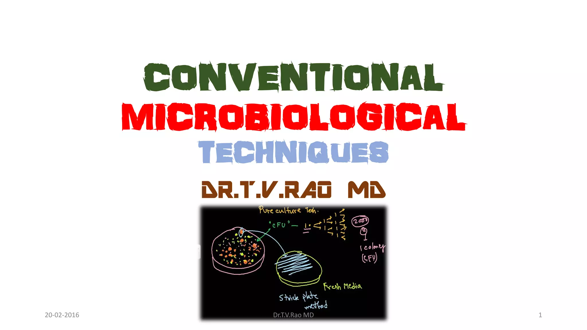
CONVENTIONAL MICROBIOLOGICAL TECHNIQUES (20)
Dr. T.V. Rao discusses what makes a good lecture. Some key points include:
- A good lecture is well-structured, engaging, and covers the essential material in a clear manner.
- Effective lecturers demonstrate expertise on the topic, use examples to illustrate concepts, and generate interest among students.
- While lecturing remains an important teaching method, especially for large classes, lecturers should aim to actively involve students through questions and other techniques.
- Preparation, clear communication, enthusiasm, and knowledge of the subject matter are hallmarks of successful lecturing. A good lecture facilitates learning while sparking students' curiosity.
The document discusses antibiotic resistance and the need for antibiotic policies in hospitals. It provides background on the development of antibiotic resistance over time. The key points are:
- Inappropriate antibiotic use promotes the spread of resistant bacteria. Hospital settings can foster drug resistance.
- An antibiotic policy aims to reduce resistance by optimizing antibiotic use and educating staff. The policy is developed with input from microbiologists, pharmacists, and clinicians.
- The hospital infection control committee implements and monitors adherence to the antibiotic policy. Continuous education is needed to ensure appropriate antibiotic prescribing.
Toxoplasmosis is caused by the parasite Toxoplasma gondii and can cause encephalitis and neurological disease in patients with low CD4 counts. It is diagnosed through imaging, blood tests, and sometimes brain biopsies. Treatment involves antiparasitic drugs and maintaining CD4 counts through antiretroviral therapy. Cryptosporidiosis is caused by Cryptosporidium parasites and causes diarrhea. It is transmitted through contaminated water or food. Microsporidiosis is caused by various protist parasites and can infect the gut or other organs. It is diagnosed through stool or tissue samples and treated with antiparasitic drugs and antiretroviral therapy. Isosporiasis is
Artificial intelligence shows promise in helping to control infectious diseases and reduce antimicrobial resistance in three key ways:
1) AI can enhance disease surveillance and early detection of outbreaks by integrating diverse data sources to identify patterns.
2) It can help optimize antimicrobial treatment by recommending personalized therapy regimens based on a patient's clinical information.
3) Over time, AI may become an indispensable public health tool by facilitating more accurate intervention strategies and optimizing resource allocation to curb disease spread.
1) Hungarian physician Ignaz Semmelweis observed higher mortality rates of women giving birth in the medical student ward compared to the midwife ward in the 1840s.
2) He discovered that the doctors in the medical student ward were coming directly from dissecting corpses to examining women without washing their hands, possibly transmitting infections.
3) Semmelweis mandated that doctors wash their hands with chlorine before examinations, which dramatically reduced the mortality rates in the medical student ward. This provided early evidence that hand hygiene reduces healthcare-associated infections.
Dr. T.V. Rao discusses causality department practices and environmental safety measures. Proper cleaning and disinfection are top priorities to prevent transmission of infectious agents and protect human safety. Dedicated cleaning practices are especially important when dealing with patients admitted with infectious diseases like diarrhea. Adherence to cleaning protocols and use of appropriate disinfectants can reduce healthcare-associated infections.
Biosecurity and infection control in hospitals aims to prevent the spread of infectious diseases. It includes proper hand hygiene, cleaning and disinfection of surfaces, use of personal protective equipment, and isolation techniques. Ensuring strict adherence to protocols through staff training and environmental monitoring is key to reducing healthcare-associated infections and protecting patients, staff, and the community.
This document discusses how microbiologists can improve clinical care through better laboratory reporting. It emphasizes providing accurate, clinically relevant results and clear interpretive comments to aid clinician decision making. Effective communication between the laboratory and clinicians is key. The document also highlights challenges such as information overload, confusion over terminology, and ensuring rapid reporting, especially for ICU patients.
This document discusses ventilator associated pneumonia (VAP), including its definition, risk factors, pathogenesis, prevention strategies, and more. Some key points:
- VAP is pneumonia that develops in intubated patients and is the leading cause of hospital-acquired infections in the ICU. The risk is 6-21 times higher for mechanically ventilated patients.
- Common risk factors for VAP include underlying illnesses, prolonged mechanical ventilation, supine position, and comorbidities like diabetes or heart failure.
- Bacteria are usually the cause, often multidrug-resistant pathogens like Pseudomonas, Klebsiella, Acinetobacter, and MRSA.
- Prevention strategies include
This document discusses hospital-acquired infections and strategies for prevention. It begins by honoring Ignaz Semmelweis, who in the 1800s established that handwashing reduced maternal mortality during childbirth by 90%. The document then discusses the nature and consequences of hospital-acquired infections. Key strategies for prevention discussed include the importance of handwashing, environmental cleaning, immunization, and establishing infection control committees and antibiotic policies. The challenges of drug resistance and maintaining standards with staff turnover are also addressed.
This document discusses biosecurity and biosafety in healthcare settings. It defines biosecurity as a strategic approach to analyzing and managing risks to human, animal, and plant life from infectious diseases. Biosafety refers to measures that reduce exposure to potentially infectious materials. The document outlines various infection control methods used in hospitals including standard precautions, hygiene practices like hand washing, and managing nosocomial infections and needlestick injuries. It emphasizes the importance of education and surveillance to improve patient safety.
Artificial intelligence has the potential to significantly impact the practice of medicine. It is being used in areas like disease diagnosis using machine learning models, personalized treatment through precision medicine, and providing virtual assistants that can answer patient questions. AI also has benefits such as improving patient safety by reducing errors, lowering healthcare costs, and increasing access to care through tools like chatbots. However, medical professionals need more education on AI applications and their ethical use to ensure they improve patient outcomes.
The document discusses MRSA (methicillin-resistant Staphylococcus aureus), including what it is, how it develops resistance, types of infections it causes, risk factors, screening and testing methods, and prevention strategies. MRSA is a strain of staph bacteria that is resistant to certain antibiotics like methicillin and oxacillin. Screening high-risk patients and implementing good hand hygiene are effective ways to control the spread of MRSA infections in healthcare settings.
This document discusses the history and methods of sterilization and disinfection. It begins with a brief history of sterilization dating back to the invention of the autoclave in 1862. It then covers terminology related to sterilization and discusses various sterilization methods including physical methods like heat, filtration, and irradiation as well as chemical methods. Factors that influence the efficacy of sterilization methods are also examined. The document provides an overview of the development and principles of sterilization.
This document discusses antimicrobial stewardship and the importance of appropriate antibiotic usage. It notes that nearly half of hospitalized patients receive antimicrobial agents. However, there has been misuse of antibiotics through treating trivial infections, commercial pressures, and a lack of understanding of antibiotic principles. This has led to a rise in antibiotic-resistant bacteria. The document advocates for antimicrobial stewardship programs in hospitals to optimize clinical outcomes while reducing unintended consequences of antibiotic usage like toxicity, resistance, and costs. Such programs involve formulary restrictions, guidelines, education, and prospective audits to ensure appropriate antibiotic selection and usage.
Cephalosporins are a class of antibiotics derived from the fungus Cephalosporium. The first generation was introduced in 1964 and provided activity against gram-positive cocci. Subsequent generations have increasingly broader coverage of gram-negative organisms. Mechanisms of resistance include beta-lactamase production and changes to penicillin-binding proteins. Later generations are used for serious hospital-acquired infections and as drugs of last resort for pathogens like Salmonella.
Coxsackieviruses were discovered in 1948-49 in Albany, New York and were named after the town of Coxsackie where samples were originally obtained. They belong to the Picornaviridae family and Enterovirus genus which also includes poliovirus and echovirus. Coxsackieviruses are divided into two groups - A and B - based on their pathogenicity in mice. Group A causes myositis while Group B causes muscle and neuronal tissue damage. Common diseases include hand-foot-and-mouth disease, herpangina, and myocarditis. Transmission is usually via the fecal-oral route. While there is no vaccine, treatment involves rest, fluids, and
This document discusses hospital-associated infections (HAIs), also known as nosocomial infections. It covers topics such as the history of infection control efforts dating back to Ignaz Semmelweiss, the impact of HAIs on patients and hospitals, common causes of HAIs, and strategies for prevention and control of infections. This includes the importance of hand hygiene, infection control committees and policies, and guidelines for reducing various device- and procedure-related infections.
The document discusses Enterobacteriaceae, a family of Gram-negative bacteria commonly found in the intestines. It describes their characteristics, classification, and important pathogenic members. Key points include:
- Enterobacteriaceae are facultative anaerobes that ferment glucose and reduce nitrates. They include both commensals and pathogens.
- Major pathogenic genera include Escherichia, Klebsiella, Salmonella, Shigella, and Proteus.
- E. coli is further classified based on virulence factors and infections caused, including gastroenteritis from enterotoxigenic, enteropathogenic, enterohemorrhagic, and enteroaggregative strains.
The document discusses antibiotics and the growing problem of antibiotic resistance. It notes that while antibiotics revolutionized medicine in the 1940s, overuse and misuse has led to more antibiotic-resistant bacteria. It describes how antibiotic resistance develops and spreads when bacteria are exposed to sub-lethal antibiotic levels. The document advocates judicious antibiotic use in humans and animals to help reduce resistance and prolong the usefulness of current antibiotics, as no new classes have been introduced in decades. It also supports documenting antibiotic susceptibility data using the WHONET program to help monitor local and global resistance trends.
More from Society for Microbiology and Infection care (20)
Hemodialysis: Chapter 8, Complications During Hemodialysis, Part 2 - Dr.GawadNephroTube - Dr.Gawad
- Video recording of this lecture in English language: https://youtu.be/FHV_jNJUt3Y
- Video recording of this lecture in Arabic language: https://youtu.be/D5kYfTMFA8E
- Link to download the book free: https://nephrotube.blogspot.com/p/nephrotube-nephrology-books.html
- Link to NephroTube website: www.NephroTube.com
- Link to NephroTube social media accounts: https://nephrotube.blogspot.com/p/join-nephrotube-on-social-media.html
Hemodialysis: Chapter 8, Complications During Hemodialysis, Part 3 - Dr.GawadNephroTube - Dr.Gawad
- Video recording of this lecture in English language: https://youtu.be/pCU7Plqbo-E
- Video recording of this lecture in Arabic language: https://youtu.be/kbDs1uaeyyo
- Link to download the book free: https://nephrotube.blogspot.com/p/nephrotube-nephrology-books.html
- Link to NephroTube website: www.NephroTube.com
- Link to NephroTube social media accounts: https://nephrotube.blogspot.com/p/join-nephrotube-on-social-media.html
Ontotext’s Clinical Trials Eligibility Design Assistant helps with one of the most challenging tasks in study design: selecting the proper patient population.
Hepatocarcinoma today between guidelines and medical therapy. The role of sur...Gian Luca Grazi
Today more than ever, hepatocellular carcinoma therapy is experiencing profound and substantial changes.
The association atezolizumab (ATEZO) plus bevacizumab (BEVA) has demonstrated its effectiveness in the post-operative treatment of patients, improving the results that can be achieved with liver resections. This after the failure of the use of sorafenib in the already historic STORM study.
On the other hand, the prognostic classification of BCLC is now widely questioned. It is now well recognized that the indications for surgery for patients with hepatocellular carcinoma are certainly narrow in BCLC and no longer reflect what is common everyday clinical practice.
Today, the concept of multiparametric therapeutic hierarchy, which makes the management of patients with hepatocellular carcinoma much more flexible and allows the best therapy for the individual patient to be identified based on their clinical characteristics, is gaining more and more importance.
The presentation traces these profound changes that are taking place in recent years and offers a modern vision of the management of patients with hepatocellular carcinoma.
A comparative study on uroculturome antimicrobial susceptibility in apparentl...Bhoj Raj Singh
The uroculturome indicates the profile of culturable microbes inhabiting the urinary tract, and it is often required to do a urine culture to find an effective antimicrobial to treat UTIs. This study targeted to understand the profile of culturable pathogens in the urine of apparently healthy (128) and humans with clinical UTIs (161). In urine samples from UTI cases, microbial counts were 1.2×10⁴ ± 6.02×10³ colony-forming units (cfu)/ mL, while in urine samples from apparently healthy humans, the average count was 3.33± 1.34×10³ cfu/ mL. In eight samples (six from UTI cases and two from apparently healthy people) of urine, Candida (C. albicans 3, C. catenulata 1, C. krusei 1, C. tropicalis 1, C. parapsiplosis 1, C. gulliermondii 1) and Rhizopus species (1) were detected. Candida krusei was detected only in a single urine sample from a healthy person and C. albicans was detected both in urine of healthy and clinical UTI cases. Fungal strains were always detected with one or more types of bacteria. Gram-positive bacteria were more commonly (OR, 1.98; CI99, 1.01-3.87) detected in urine samples of apparently healthy humans, and Gram -ve bacteria (OR, 2.74; CI99, 1.44-5.23) in urines of UTI cases. From urine samples of 161 UTI cases, a total of 90 different types of microbes were detected and, 73 samples had only a single type of bacteria. In contrast, 49, 29, 3, 4, 1, and 2 samples had 2, 3, 4, 5, 6 and 7 types of bacteria, respectively. The most common bacteria detected in urine of UTI cases was Escherichia coli detected in 52 samples, in 20 cases as the single type of bacteria, other 34 types of bacteria were detected in pure form in 53 cases. From 128 urine samples of apparently healthy people, 88 types of microbes were detected either singly or in association with others, from 64 urine samples only a single type of bacteria was detected while 34, 13, 3, 11, 2 and 1 samples yielded 2, 3, 4, 5, 6 and seven types of microbes, respectively. In the urine of apparently healthy humans too, E. coli was the most common bacteria, detected in pure culture from 10 samples followed by Staphylococcus haemolyticus (9), S. intermedius (5), and S. aureus (5), and similar types of bacteria also dominated in cases of mixed occurrence, E. coli was detected in 26, S. aureus in 22 and S. haemolyticus in 19 urine samples, respectively. Gram +ve bacteria isolated from urine samples' irrespective of health status were more often (p, <0.01) resistant than Gram -ve bacteria to ajowan oil, holy basil oil, cinnamaldehyde, and cinnamon oil, but more susceptible to sandalwood oil (p, <0.01). However, for antibiotics, Gram +ve were more often susceptible than Gram -ve bacteria to cephalosporins, doxycycline, and nitrofurantoin. The study concludes that to understand the role of good and bad bacteria in the urinary tract microbiome more targeted studies are needed to discern the isolates at the pathotype level.
High Profile"*Call "*Girls in Kolkata ))86-075-754-83(( "*Call "*Girls in Kol...Nisha Malik Chaudhary
High Profile "*Call "*Girls in Kolkata ))86-075-754-83(( "*Call "*Girls in Kolkata Available
Kolkata "Call "Girls 74046-34175 "Call "Girl Number in Kolkata | A nutshell review for Hot "Call "Girls in Kolkata . MY experience was superb with them this is the only recommended "Call "Girls service in Kolkata "Call "Girls and again then Russian. so overall my practice was magnificent. The price is also moderate per hour. The plus point is the "Girl comes instantly to your lo"Cation doesn't matter you are in Bur Kolkata or al Nahda or Kolkata or any area she comes undeviatingly to your hotel room. Definitely recommend the "Call "Girls agency. A nutshell review for Hot "Call "Girls in Kolkata . MY experience was superb with them this is the only recommended "Call "Girls service in Kolkata with verified "Call "Girls . I am using their services from past 6 months they never ever disappointed me in any way. Let's just say if i asked them to provide me russian "Call "Girls they fulfilled my request or even beautiful "Call "Girls or indian "Call "Girls in Kolkata . They have their owen drivers who brings the "Call "Girls in less time in any area of Kolkata like bur Kolkata marina or jumeirah or even in jebel ali as well. I'm writing here everything after experience their services in all conditions.
Exploring Alternatives- Why Laparoscopy Isn't Always Best for Hydrosalpinx.pptxFFragrant
Not all women with hydrosalpinx should choose laparoscopy. Natural medicine Fuyan Pill can also be a nice option for patients, especially when they have fertility needs.
Ventilation Perfusion Ratio, Physiological dead space and physiological shuntMedicoseAcademics
In this insightful lecture, Dr. Faiza, an esteemed Assistant Professor of Physiology, delves into the essential concept of the ventilation-perfusion ratio (V˙/Q˙), which is fundamental to understanding pulmonary physiology. Dr. Faiza brings a wealth of knowledge and experience to the table, with qualifications including MBBS, FCPS in Physiology, and multiple postgraduate degrees in public health and healthcare education.
The lecture begins by laying the groundwork with basic concepts, explaining the definitions of ventilation (V˙) and perfusion (Q˙), and highlighting the significance of the ventilation-perfusion ratio (V˙/Q˙). Dr. Faiza explains the normal value of this ratio and its critical role in ensuring efficient gas exchange in the lungs.
Next, the discussion moves to the impact of different V˙/Q˙ ratios on alveolar gas concentrations. Participants will learn how a normal, zero, or infinite V˙/Q˙ ratio affects the partial pressures of oxygen and carbon dioxide in the alveoli. Dr. Faiza provides a detailed comparison of alveolar gas concentrations in these varying scenarios, offering a clear understanding of the physiological changes that occur.
The lecture also covers the concepts of physiological shunt and dead space. Dr. Faiza defines physiological shunt and explains its causes and effects on gas exchange, distinguishing it from anatomical dead space. She also discusses physiological dead space in detail, including how it is calculated using the Bohr equation. The components and significance of the Bohr equation are thoroughly explained, and practical examples of its application are provided.
Further, the lecture examines the variations in V˙/Q˙ ratios in different regions of the lung and under different conditions, such as lying versus supine and resting versus exercise. Dr. Faiza analyzes how these variations affect pulmonary function and discusses the abnormal V˙/Q˙ ratios seen in chronic obstructive lung disease (COPD) and their clinical implications.
Finally, Dr. Faiza explores the clinical implications of abnormal V˙/Q˙ ratios. She identifies clinical conditions associated with these abnormalities, such as COPD and emphysema, and discusses the physiological and clinical consequences on respiratory function. The lecture emphasizes the importance of understanding these concepts for medical professionals and students, highlighting their relevance in diagnosing and managing respiratory conditions.
This comprehensive lecture provides valuable insights for medical students, healthcare professionals, and anyone interested in respiratory physiology. Participants will gain a deep understanding of how ventilation and perfusion work together to optimize gas exchange in the lungs and how deviations from the norm can lead to significant clinical issues.
Descoperă Bucuria Vieții Sănătoase cu Jurnalul Fericirii Life Care - Iulie 2024!
Gata să te bucuri de o vară vibrantă și plină de energie? Life Care îți vine în ajutor cu Jurnalul Fericirii din Iulie 2024, un ghid complet pentru o viață armonioasă și echilibrată.
Pe parcursul a cateva de pagini pline de informații utile și inspirație, vei descoperi:
Sfaturi practice pentru o alimentație sănătoasă:
Rețete delicioase și ușor de preparat: Bucură-te de preparate gustoase și nutritive, perfecte pentru zilele călduroase de vară.
Recomandări pentru o alimentație echilibrată: Asigură-ți aportul necesar de nutrienți esențiali pentru un organism sănătos și plin de vitalitate.
Sfaturi pentru alegeri alimentare inteligente: Învață cum să faci cumpărături sănătoase și să eviți tentațiile nesănătoase.
Trucuri pentru un stil de viață activ:
Rutine de exerciții fizice adaptate nevoilor tale: Găsește antrenamente potrivite pentru a te menține în formă și energic pe tot parcursul verii.
Idei de activități în aer liber: Descoperă modalități distractive de a te bucura de vremea frumoasă și de a petrece timp de calitate cu cei dragi.
Sfaturi pentru un somn odihnitor: Asigură-ți un somn profund și reparator pentru a te trezi revigorat și pregătit pentru o nouă zi.
Sfaturi pentru o stare de bine mentală:
Tehnici de relaxare și gestionare a stresului: Învață cum să te relaxezi și să faci față provocărilor zilnice cu mai multă ușurință.
Sfaturi pentru cultivarea optimismului și a gândirii pozitive: Descoperă cum să abordezi viața cu o perspectivă optimistă și să atragi mai multă bucurie în ea.
Recomandări pentru a te conecta cu natura: Bucură-te de beneficiile naturii asupra stării tale mentale și emoționale.
Bonus:
Oferte exclusive la produsele Life Care: Beneficiază de reduceri și promoții speciale la o gamă largă de produse pentru o viață sănătoasă.
Concursuri și premii: Participă la concursuri distractive și câștigă premii valoroase.
Jurnalul Fericirii Life Care - Iulie 2024 este mai mult decât o simplă revistă. Este un ghid complet și personalizat pentru a te ajuta să obții o viață mai sănătoasă, mai fericită și mai plină de satisfacții.
Nu rata această șansă de a te bucura de vară la maximum! Descoperă Jurnalul Fericirii Life Care - Iulie 2024 astăzi!
Comandă-ți exemplarul acum și fă un pas important către o viață mai bună!
#JurnalulFericirii #LifeCare #Iulie2024 #ViataSanatoasa #Bunastare #Fericire #Oferte #Concursuri #Premii
Coronary Circulation and Ischemic Heart Disease_AntiCopy.pdfMedicoseAcademics
In this lecture, we delve into the intricate anatomy and physiology of the coronary blood supply, a crucial aspect of cardiac function. We begin by examining the physiological anatomy of the coronary arteries, which lie on the heart's surface and penetrate the cardiac muscle mass to supply essential nutrients. Notably, only the innermost layer of the endocardial surface receives direct nourishment from the blood within the cardiac chambers.
We then explore the specifics of coronary circulation, including the dynamics of blood flow at rest and during strenuous activity. The impact of cardiac muscle compression on coronary blood flow, particularly during systole and diastole, is discussed, highlighting why this phenomenon is more pronounced in the left ventricle than the right.
Regulation of coronary circulation is a complex process influenced by autonomic and local metabolic factors. We discuss the roles of sympathetic and parasympathetic nerves, emphasizing the dominance of local metabolic factors such as hypoxia and adenosine in coronary vasodilation. Concepts like autoregulation, active hyperemia, and reactive hyperemia are explained to illustrate how the heart adjusts blood flow to meet varying oxygen demands.
Ischemic heart disease is a major focus, with an exploration of acute coronary artery occlusion, myocardial infarction, and subsequent physiological changes. The lecture covers the progression from acute occlusion to infarction, the body's compensatory mechanisms, and the potential complications leading to death, such as cardiac failure, pulmonary edema, fibrillation, and cardiac rupture.
We also examine coronary steal syndrome, a condition where increased cardiac activity diverts blood flow away from ischemic areas, exacerbating the condition. The long-term impact of myocardial infarction on cardiac reserve is discussed, showing how the heart's capacity to handle increased workloads is significantly reduced.
Angina pectoris, a common manifestation of ischemic heart disease, is analyzed in terms of its causes, presentation, and referred pain patterns. We identify factors that exacerbate anginal pain and discuss both medical and surgical treatment options.
Finally, the lecture includes a case study to apply theoretical knowledge to a practical scenario, helping students understand the real-world implications of coronary circulation and ischemic heart disease. The role of biochemical factors in cardiac pain and the interpretation of ECG changes in myocardial infarction are also covered.
4. A great quotes by Ronald Ross
• I have failed in finding parasites
in mosquitoes fed on malaria
patients, but perhaps I am not
using the proper kind of
mosquito
• “The screws of my microscope
were rusted with sweat from my
forehead and hands, and its last
remaining eye-piece was cracked
20-02-2016 Dr.T.V.Rao MD 4
5. Beginning of Diagnostic Microbiology
•In the late 1800s, the realization that
identifiable microbes caused specific
diseases led to pathogens Specific medical
diagnosis. Although the time honoured
techniques of growing bacteria in broth or
solid cultures and staining and examining
them under microscopes are still important
today20-02-2016 Dr.T.V.Rao MD 5
6. A GREAT QUESTION TODAY HOW
KEEN WE ARE WITH THE WORK
20-02-2016 Dr.T.V.Rao MD 6
8. The Real Good of the Past in
Diagnostic Microbiology
• In the good old days, the
microbiology laboratory used
to be a labour intensive
place equipped with
incubators and microscopes.
Microbiologists were patient
scientists waiting at least 24
hours before their isolated
cultures were grown enough
for identification
20-02-2016 Dr.T.V.Rao MD 8
9. Where we stand Today
• Most neglected and least
invested specialty
• Reasons can be many
20-02-2016 Dr.T.V.Rao MD 9
11. What is a Technique
A way of carrying out
a particular task,
especially the
execution or
performance of an
artistic work or a
scientific procedure
20-02-2016 Dr.T.V.Rao MD 11
12. American Society for Microbiology encourages Artistic and
Conventional Techniques on Streaking and Culturing
20-02-2016 Dr.T.V.Rao MD 12
13. A MATTER OF CONFLICT
WHO WILL COLLECT THE SPECIMENS
• Proper specimen collection,
container labelling, and
culture requests are the
responsibility of the
ordering physician.
Technologists in the Clinical
Microbiology Laboratory will
be familiar with specimens
of choice and proper
collection techniques
20-02-2016 Dr.T.V.Rao MD 13
14. 14
SPECIMEN COLLECTION
whose duty it is ?
The specimen is the beginning. All
diagnostic information from the laboratory
depends upon the knowledge by which
specimens are chosen and the care with
which they are collected and transported.
—Cynthia A. Needham
20-02-2016 Dr.T.V.Rao MD
15. If you accept the Truth many Suboptimal
Specimens are Processed
• I should share my
experience the ideal
collecting of a sample
remain with greater
challenges, we many
times receive
suboptimal sample for
processing, few posses
courage to reject, it just
goes
20-02-2016 Dr.T.V.Rao MD 15
16. LABORATORY HANDLES THE SPECIMENS
• The technologist in the
laboratory will directly
handle specimens of
clinical and
environmental source
which are received from
the Postal Service or
hand carried to the
laboratory.
20-02-2016 Dr.T.V.Rao MD 16
17. Are we Collecting the Right specimens
• Select an appropriate
sites or organs for
sampling.
• Use sterile equipment
for sampling.
• Labelling the sample
• Keep and transfer the
sample inappropriate
medium and condition.20-02-2016 Dr.T.V.Rao MD 17
18. Microbiological techniques
• Bacteria will grow on
practically any source of
organic food which provides
carbon compounds to be
respired for energy, and
nitrogen compounds to be
incorporated into proteins
for growth. These
substances are normally
provided dissolved in water.
20-02-2016 Dr.T.V.Rao MD 18
20. General View on the Parameters Used in the Process of
Microorganism
Identification
• Before one can proceed
to identify a
microorganism, the
characteristics of that
organism have to be
determined in details.
The major characteristics
which are observed
20-02-2016 Dr.T.V.Rao MD 20
21. Gram Staining continues to be Most rapid method
to diagnose
• The rapid Gram stain evaluation is
reliable, easy to perform, and well
suited for the routine clinical
laboratory
• Eg – CSF examination in
emergencies
• Sputum in case of acute
pneumonias
• Bacterial Vaginitis
• Any other transudates
• Pleural fluid
• Pericardial fluids
20-02-2016 Dr.T.V.Rao MD 21
22. Molecular Biology in Infancy
• Molecular biology
techniques promise to
revolutionize the
diagnosis of infectious
disease—to date a
promise still in its
infancy
20-02-2016 Dr.T.V.Rao MD 22
23. Laboratory Medicine Under threat
Microbiology is No exception
•Great questions to many
Microbiologists
How much Time we are spending in the
Laboratory
Who are doing the Bench work
Are laboratories equipped with minimal
requirements of infrastructure and Biosafety?20-02-2016 Dr.T.V.Rao MD 23
25. Use a bright field light microscope
• Use a bright field light
microscope to view and
interpret slides, including
• 1. correctly setting up and
focusing the microscope
• 2. proper handling, cleaning, and
storage of the microscope
• 3. correct use of all lenses
• 4. recording microscopic
observations
20-02-2016 Dr.T.V.Rao MD 25
26. Properly prepare slides
• Properly prepare slides for
microbiological examination,
including
• A. cleaning and disposing of
slides
• B. preparing smears from solid
and liquid cultures
• C. performing wet mount and/or
hanging drop preparations
• D. performing Gram stains
20-02-2016 Dr.T.V.Rao MD 26
27. Properly use aseptic techniques
• Properly use aseptic techniques
for the transfer and handling of
microorganisms and
instruments, including
• A. sterilizing and maintaining
sterility of transfer instruments
• B. performing aseptic transfer
• C. obtaining microbial samples
20-02-2016 Dr.T.V.Rao MD 27
28. Use appropriate microbiological Media
and Test Systems
• A. Isolating colonies
and/or plaques
• B. Maintaining pure
cultures
• C. Using biochemical test
media
• C. Accurately recording
macroscopic
observations20-02-2016 Dr.T.V.Rao MD 28
29. Use standard microbiology laboratory
equipment correctly
• A. using the standard
metric system for
weights, lengths,
diameters, and volumes
• B. lighting and adjusting a
laboratory burner
• C. using an incubator
right Temperature to be
monitored20-02-2016 Dr.T.V.Rao MD 29
30. Streaking is the Beginning of learning
Alphabets in Microbiology
20-02-2016 Dr.T.V.Rao MD 30
31. Streaking the Urine specimens is the best way
to learn the matters with precision
20-02-2016 Dr.T.V.Rao MD 31
32. Streaking is Key to success in Separation of
Pathogens from Commensals
20-02-2016 Dr.T.V.Rao MD 32
33. KNOW THE MICROBES BEFORE YOU
PROCEED
• The cultural characteristics of a microorganism usually
vary depending on the media used and many other
factors. Some experienced microbiologists could have
a good guess about the identity of a microorganism
just by its cultural characteristics, but this was proven
to be a bad technique. Students as well as
microbiologists are advised to follow strict procedures
for the identification of isolates from clinical
specimens.
20-02-2016 Dr.T.V.Rao MD 33
35. General View on the Parameters Used in the Process of
Microorganism
Identification
• Before one can proceed to identify a microorganism, the
characteristics of that organism have to be determined in
details In clinical terms, it is the shape, size, colour, elevation
and other characteristics of the colony formed on the culture
plate. In taxonomy, it includes the nutrient requirements for
the growth of the organism and the physical factors such as
temperature, pH and the incubation period. These factors
are used to identify certain pathogenic species but less
commonly used in routine procedures
20-02-2016 Dr.T.V.Rao MD 35
36. Is our experience Matches Scientific Approach
• Some experienced
microbiologists could have a
good guess about the identity of
a microorganism just by its
cultural characteristics, but this
was proven to be a bad
technique. Students as well as
microbiologists are advised to
follow strict procedures for the
identification of isolates from
clinical specimens.
20-02-2016 Dr.T.V.Rao MD 36
37. BIOCHEMICAL CHARACTERISTICS
• Frequently, the identity of a species requires detailed
knowledge of its biochemical activities, since other
characteristics are not sufficiently distinctive or differential.
For example, the bacterium Escherichia coli, a normal
inhabitant of our intestinal tract, is indistinguishable
microscopically from Salmonella typhi, the bacterium that
causes typhoid fever. However, if these two bacteria are
examined for their metabolic (or biochemical)
characteristics, they are found to be very different and
distinguishable on this basis.
20-02-2016 Dr.T.V.Rao MD 37
38. Morphology and Staining
• This includes the
microscopic appearance of
a stained preparation of the
organism. Useful
information to be taken into
account, are the size of the
individual cells, cell shape
and arrangement and
staining reaction if
differential staining
procedures is used.20-02-2016 Dr.T.V.Rao MD 38
39. TILL TO DATE THERE IS NO FASTER
METHOD AS GRAM’S STAINING
• Some laboratories which
have a little facility could
give the report of a
microbiological
examination of a clinical
specimen just by stating
their morphological
characteristics and the
sensitivity testing results
20-02-2016 Dr.T.V.Rao MD 39
40. LABORATORY HANDLES THE SPECIMENS
• The technologist in the
laboratory will directly
handle specimens of
clinical and
environmental source
which are received from
the Postal Service or
hand carried to the
laboratory.
20-02-2016 Dr.T.V.Rao MD 40
41. Gram staining most Rapid method as the
Situation warrants
• Look up reference images. If
you are not certain what a
bacteria is, look through a
collection of reference
images, sorted by shape and
result of the gram stain. You
can find databases online at
the National Microbial
Pathogen Database
20-02-2016 Dr.T.V.Rao MD 41
42. Disadvantages of Microscopic methods
•Microscopy may suggest an etiologic agent, but
it rarely provides definitive evidence of infection
by a particular species.
• Microscopic findings regarding bacterial
morphology may be misleading, because many
species can be pleomorphic and conclusions can
be influenced by subjective interpretation of the
investigator.
20-02-2016 Dr.T.V.Rao MD 42
43. Staining is limited Sensitive
• Limited sensitivity is because a relatively large number
of microbial cells are required before they are seen
under microscopy (e.g. 104 bacterial cells/ml of fluid)
(Fredricks & Relman, 1999). Some micro-organisms
can even require appropriate stains and/or
approaches to become visible.
• Limited specificity is because our inability to speciate
micro-organisms based on their morphology and
staining patterns.
20-02-2016 Dr.T.V.Rao MD 43
44. Problems With Traditional Methods
• Cultivation-based methods insensitive for detecting some
organisms.
• Cultivation-based methods limited to pathogens with known
growth requirements.
• Poor discrimination between microbes with common
behavioural features.
• Failure to detect infections caused by uncultivated (e.g.,
novel) organisms, or organisms that fail to elicit a detectable
host immune response.
• Visual appearance of microorganisms is nonspecific.
20-02-2016 Dr.T.V.Rao MD 44
45. Problems With Traditional Methods
• Examples of Failures With Traditional Approaches
• Detection and speciation of slow-growing organisms takes
weeks
• (e.g., M. tuberculosis).
• A number of visible microorganisms cannot be cultivated
(e.g., Whipple bacillus).
• Diseases presumed to be infectious remain ill-defined with
not detected microorganism (e.g., abrupt fever after tick
bite).
20-02-2016 Dr.T.V.Rao MD 45
46. Culture of fungi
• Fungi specimens
obtained from non sterile
sites must be inoculated
onto media containing
antibacterial agents.
Specimens should be
allowed to grow for 4 wk
before being discarded.
20-02-2016 Dr.T.V.Rao MD 46
47. Use of Physical Separation Procedures
Streak Plate technique
•As we have seen in
previous labs,
single colonies
may be achieved
by using the streak
plate technique
20-02-2016 Dr.T.V.Rao MD 47
49. Most of us Miss the Parasites
•Parasites cause hidden epidemics, many practitioners
do not even think
•Microbiologists evaluate for Bacteriological examination
can miss the Parasites and Fungus
•Still our society there sare several parasitic infections
prevails
•Simpler techniques are missed as stool examination and
entrusted with most of the times to the technicians
without much understanding20-02-2016 Dr.T.V.Rao MD 49
50. Can we diagnose the stool parasites by
conventional methods with accuracy
•Microsporidia
•Isospora Belli &
Cyclospora
•Cryptosporidium
20-02-2016 Dr.T.V.Rao MD 50
51. Parasitology Testing Laboratory Services
• This test evaluates stool for presence of parasites and levels of
beneficial flora, imbalanced flora, possible pathogenic
bacteria and possible fungal pathogens. This Comprehensive
Parasitology Profile uses the most technologically advanced
procedures to accurately identify a wide range of protozoal
parasites, including amoebae, flagellates, ciliates, coccidia and
microsporidia. This stool test can help reveal hidden causes
behind acute or chronic conditions that develop from
parasitic infection or dysbiosis.
20-02-2016 Dr.T.V.Rao MD 51
52. Empowering conventional techniques with
modern technology
• Specimens are carefully analysed by highly trained
technicians using computer-enhanced video
microscopy, new staining procedures, and advanced
immunoassay techniques. These accurate detection
methods allow for increased detection rates,
increasing the awareness of the important
relationship between parasitic infection and a broad
spectrum of illnesses and diseases
20-02-2016 Dr.T.V.Rao MD 52
53. IS THE CONVENTIONAL METHODS CONVINCING
THE CLINICIANS YES / NO
•I am confident it is
certainly no many
times tests are
ordered without
forethought on the
existing the clinical
diagnosis and it is just
fishing in troubled
water,20-02-2016 Dr.T.V.Rao MD 53
54. Not a one step Diagnosis
•Even Today Many
Clinicians think
Microbiologist
diagnosis is one step
diagnosis as in
Biochemistry as we
many steps to be
perform coming to
minimal conclusions20-02-2016 Dr.T.V.Rao MD 54
55. MOVING TO FEATURE NEEDS
• In the get-it-done-yesterday environment Hospitals
and Microbiology laboratories are finding that the
traditional microbiological methodologies and
especially sending microbiological samples to outside
labs cost them time, money and opportunities
Internalizing the microbiology testing and especially
adopting rapid microbiological methods (RMM) can
significantly speed up the time to results from 7-10
days to 24-48 hours.
20-02-2016 Dr.T.V.Rao MD 55
56. Limitations of Conventional Microbiological Testing
• Conventional microbiological
methods have well-known
inherent limitations. These
include small test sample
volumes, prolonged incubation
periods, incompatibilities with
membrane filtration, and
ambiguity associated with using
turbidity as a detection endpoint
20-02-2016 Dr.T.V.Rao MD 56
57. India can face many challenges
• The increase in worldwide travel,
coupled with increasing
immigration into the India,
contributes to the spread and
incidence of parasitic infections.
In addition, parasitic infections
are commonly transmitted
through fecally contaminated
food, water, or other materials
within this country.
20-02-2016 Dr.T.V.Rao MD 57
58. Conventional techniques of Malaria
• Current diagnostic methods for
malaria include clinical diagnosis,
microscopy, serology, molecular
diagnosis, and antibody detection.
Giemsa staining of the peripheral
blood smear is the gold standard
for the diagnosis of malaria, but
also has limitations. Using
microscopy as the only means of
diagnosing malaria can lead to
false negatives. Another diagnostic
tool is Rapid Diagnostic Tests or
RDTs also known as dipstick assays.
It
20-02-2016 Dr.T.V.Rao MD 58
59. Gold Standard for diagnosis of Malaria
• The gold standard for the diagnosis
of malaria is the Giemsa staining of
the peripheral blood smear(Thick
and thin smear), but its also has its
limitations. However, even under
optimal conditions the sensitivity
of microscopy is only about twenty
parasites/μl of blood, and
subjective interpretation and
reader errors further reduce the
accuracy of diagnosis.
20-02-2016 Dr.T.V.Rao MD 59
60. Human dedication matters
• Conventional microscopic
examination of peripheral thick
and thin blood smears remains
the gold standard for malaria
diagnosis. Although this method
requires a trained microscopist,
and sensitivity and specificity
vary compared with recent
technical advances, it is
inexpensive and reliable.
20-02-2016 Dr.T.V.Rao MD 60
61. Medical Microbiologists losing the working
opportunities
• While Rapid Microbiological
Methods (RMM) offer high
degree of automation,
significant reduction in time
to results, faster product
release, ability to employ
non-microbiologists to
operate the system, and
improved control;
20-02-2016 Dr.T.V.Rao MD 61
62. My Dear Young Microbiologists
• Most people want to do excellent work. Apathy becomes a problem
when team members feel there is no solution or they have no voice
• I wish you are the future leaders in Microbiology for next 40 years
hope you all wish for change to live in comfort,
• Assess your role
• Purpose of the speciality
• Try Impress others with hard work and sincerity
• If Medical People do not wish to work some body take you place ?
• NEXT WHAT ?
20-02-2016 Dr.T.V.Rao MD 62
65. CDC helps in Digital diagnosis
➲The CDC now offers tele diagnosis to help laboratories diagnose
malaria and other parasitic diseases. When laboratories are not
certain about identifying parasites on a slide, they can e-mail to
the CDC images of the suspected parasites. Experts then review
the images and discuss findings with the submitting lab within
only a few hours, allowing near real-time diagnosis as well as an
opportunity for training in microscopic diagnosis.
20-02-2016 Dr.T.V.Rao MD 65
66. A long parasite of 15 cm was extracted without
damage to the physical structure
•An approximately 15
cm long filamentous
macroscopic
parasite was
extracted sent intact
in Normal saline, to
Microbiology
20-02-2016 Dr.T.V.Rao MD 66
68. As reported from CDC – Atlanta USA
• Based on the images, we agree this
is a female Dirofilaria (possibly D.
repens in India), as indicated by tall,
polymyarian musculature, external
cuticular ridges, and paired
reproductive tubes.
20-02-2016 Dr.T.V.Rao MD 68
69. World First for Malaria - mobile phone diagnosis
now available
• xRapid is a world first in
mobile health, providing
automatic diagnosis of
malaria via an iPhone app. It
is the first commercially
available mobile app that
has the functionality to
quickly and accurately
diagnose a major disease.
20-02-2016 Dr.T.V.Rao MD 69
70. ARE WE READY FOR CHANGE TO
AUTOMATION ?
20-02-2016 Dr.T.V.Rao MD 70
71. THE FUTURE OF DIAGNOSTIC MICROBIOLOGY
IS CHANGING
• The physical structure of laboratories, staffing
patterns, work flow, and turnaround time have all
been profoundly influenced by technical advances.
The implementation of nucleic acid amplification-
based molecular techniques provides complementary,
rapid, and on-demand diagnosis services. These
changes will continue, and lead diagnostic
microbiology inevitably to a modern discipline, which
can face many challenges in the future.
20-02-2016 Dr.T.V.Rao MD 71
72. TODAYS PROBLEM WITH DIAGNOSTIC
MICROBIOLOGY
• We Teach More than what
we see, and diagnose
• WE Talk More ?
• We discuss more ?
• However we do less ?
• 1Bench work
2 Fail to improve the
laboratory skills ?
20-02-2016 Dr.T.V.Rao MD 72
73. Laboratories should progress with scientific
Developments
•Modern science is fast-moving, and no
laboratory can exist for long with a program
based on old facilities. Innovation and renewal
are required to keep a laboratory on the
frontiers of science.
• Burton Richter
20-02-2016 Dr.T.V.Rao MD 73
74. Wish to be A Better Microbiologist
• A microbiologist needs to be
both brilliant and methodical. An
ability to think critically and
analytically is a prerequisite, as
is an advanced understanding
and knowledge of computers.
• Microbiologists must be
experts at working with
statistics and must stay
abreast of developments in
statistical techniques
20-02-2016 Dr.T.V.Rao MD 74
75. How we can improve our Diagnostic
Microbiology
20-02-2016 Dr.T.V.Rao MD 75