Definition, factors affecting electrophoresis, classification of electrophoresis in general, Iso-electric focusing in detail, IEF and its types (based on ampholytes), step wise procedure of IEF process, Problems involved and their remedies, Capillary iso electric focusing and its types, detection of analytes explained in animation (so watch it in slide show mode), advantages and applications of CIEF.
This document discusses electrophoresis, which is the migration of charged particles through a liquid medium under the influence of an electric field. It defines key terms and describes the theory behind electrophoresis, factors that influence particle migration rates, and different electrophoresis techniques. Some main techniques covered are agarose gel electrophoresis, polyacrylamide gel electrophoresis, isoelectric focusing, and two-dimensional electrophoresis. Troubleshooting tips for common issues are also provided.
Hemolytic anemias share the following features:
A shortened red cell life span below the normal 120 days
Elevated erythropoietin levels and a compensatory increase in erythropoiesis
Accumulation of hemoglobin degradation products that are created as part of the process of red cell hemolysis
Electrophoresis is a technique used to separate charged particles such as proteins, nucleic acids, and other molecules based on their size and charge. It works by applying an electric field to encourage the particles to migrate through a gel or other medium at different rates. The document discusses the components, principles, and applications of electrophoresis, including different types of gels, buffers used, and methods of visualization. It is commonly used in fields like molecular biology, genetics, and forensics.
Serum protein electrophoresis & their clinical importanceDr.M.Prasad Naidu
This document discusses serum proteins and electrophoresis techniques used to analyze them. It provides details on the major serum proteins - albumin and globulins - and their functions. Electrophoresis separates proteins based on their charge and size. Several electrophoresis methods are described, including agarose gel, SDS-PAGE, and capillary electrophoresis. Factors influencing electrophoresis results and common stains used are also outlined. The document concludes with descriptions of normal and abnormal serum protein electrophoresis patterns and their clinical significance.
Zone electrophoresis is an electrophoretic technique used to separate charged particles like proteins, nucleic acids, and biopolymers. It works by migrating the charged particles through a stabilizing medium like paper, agarose gel, or polyacrylamide gel under the influence of an electric field. The separated components form discrete zones on the supporting medium. Common types of zone electrophoresis include paper electrophoresis, gel electrophoresis using agarose or polyacrylamide, cellulose acetate electrophoresis, and thin layer electrophoresis. Each technique has advantages and applications for separating different types of biological molecules.
Electrophoresis is a technique used to separate macromolecules like proteins and nucleic acids based on their size and charge. It works by applying an electric field to a gel or membrane support medium, causing the charged molecules to migrate at different velocities depending on their size and charge. Common support media include paper, cellulose acetate, and polyacrylamide gels. Hemoglobin electrophoresis specifically separates hemoglobin types and variants to diagnose hemoglobinopathies by running blood samples on a cellulose acetate membrane in an electric field.
Electrophoresis is a technique used to separate charged molecules such as proteins and nucleic acids. It works by applying an electric field to move these molecules through a buffer or gel based on their size and charge. There are several types of electrophoresis including gel electrophoresis, which separates molecules based on size using a gel matrix, and capillary electrophoresis, which uses narrow capillaries to achieve high resolution separations. Electrophoresis has many applications in areas like biochemistry, genetics, and medicine.
Capillary electrophoresis is a technique that uses narrow bore capillaries to separate charged molecules via electrophoretic mobility. When a voltage is applied, molecules migrate through the capillary at different rates depending on their charge and size. This allows analytes like proteins, nucleic acids, and small molecules to be separated. Key advantages are high efficiency, short analysis times, and low sample volume requirements. Common modes include capillary zone electrophoresis, capillary gel electrophoresis, and micellar electrokinetic capillary chromatography. Applications include analysis in pharmaceuticals and detection of microbial contamination.
At any given pH, molecules exist as electrically charged species that will migrate toward the cathode or anode under the influence of an electric field, depending on their net charge. Electrophoresis techniques separate these charged particles using an electric current applied across a buffer solution and supporting medium like agarose gel or polyacrylamide. The basic equipment required consists of a power supply delivering current between electrodes in an electrophoresis unit, which can perform vertical or horizontal gel separations. Proteins and other analytes are visualized after separation by staining or other detection methods. Electrophoresis has numerous applications in fields like medicine, biochemistry, and food analysis.
It contains 2-3 acetyl groups per glucose unit and its adsorption capacity is less than that of paper.
It gives sharper bands.
Provides a good background for staining glycoproteins.
ADVANTAGE:
No tailing of proteins or hydrophilic materials.
Available in wide range of particle size and layer thickness.
Give sharp bands and offer good resolution.
High voltage can be applied which will enhance the resolution.
Gel Electrophoresis (suchita rawats conflicted copy 2023-04-28) (1).pptxSuchita Rawat
Gel electrophoresis is a method used to separate biomolecules like DNA, RNA, and proteins based on their size and charge. During electrophoresis, an electric current is applied to a gel, causing charged molecules to migrate through the gel at different rates depending on factors like their mass and shape. Common types of electrophoresis include agarose gel electrophoresis to separate DNA fragments, and SDS-PAGE gel electrophoresis to separate denatured proteins by size. Gel electrophoresis is a fundamental technique used in various applications in molecular biology and biochemistry.
Protein electrophoresis is a method used to separate proteins in a blood sample using an electric current. In the procedure, the serum sample is loaded onto a gel support medium like cellulose acetate. An electric field is applied using a buffer solution, causing the proteins to migrate based on their molecular weight and charge. The proteins separate into bands representing different protein types, such as albumin, alpha 1 globulin, alpha 2 globulin, beta globulin, and gamma globulin. After the bands form, the gel is stained to visualize the proteins, then a densitometer can quantify the amount of each band. This technique allows analysis of a protein mixture and detection of abnormalities.
Thank you for the detailed presentation on electrophoresis. I appreciate you taking the time to explain the key concepts and techniques. Please let me know if you have any other questions.
This document provides an overview of electrophoresis, including:
1. Electrophoresis uses the migration of charged solutes or particles in a liquid medium under the influence of an electric field. It is widely used to separate biological molecules like proteins.
2. Particles with different charge-to-mass ratios migrate at different rates depending on factors like their net charge, size, and the pH and strength of the buffer solution. Agarose gel and polyacrylamide gel electrophoresis are commonly used techniques.
3. The general procedure involves separating the particles in an electric field, staining to visualize the bands, then detecting and quantifying the separated fractions. Automated systems now allow high-throughput processing of
This document summarizes the process of 2-D gel electrophoresis used to separate proteins. It begins with an introduction explaining that 2-D gel electrophoresis couples isoelectric focusing in the first dimension based on isoelectric point and SDS-PAGE in the second dimension based on molecular mass. The document then covers the principles, processes, and techniques involved in isoelectric focusing, SDS-PAGE, blotting, and concludes by discussing the applications and references for 2-D gel electrophoresis.
This document provides information on electrophoresis, including:
1. Electrophoresis is a technique used to separate charged particles like proteins and nucleic acids in an electric field based on their charge-to-mass ratio.
2. Agarose and polyacrylamide gels are commonly used supporting media that provide a matrix for particle separation.
3. The general procedure involves sample application, electrophoretic separation, staining to visualize bands, and detection/quantification of bands through densitometry.
Electrophoresis is the migration of charged particles through an electric field towards the electrode of opposite charge. It works by separating molecules based on their size and charge. Key factors that affect electrophoresis include the net charge and size/shape of molecules, as well as the ionic strength, pH, temperature, and type of support medium used. Common applications include separating proteins, nucleic acids, and other biological molecules to analyze samples.
Similar to 
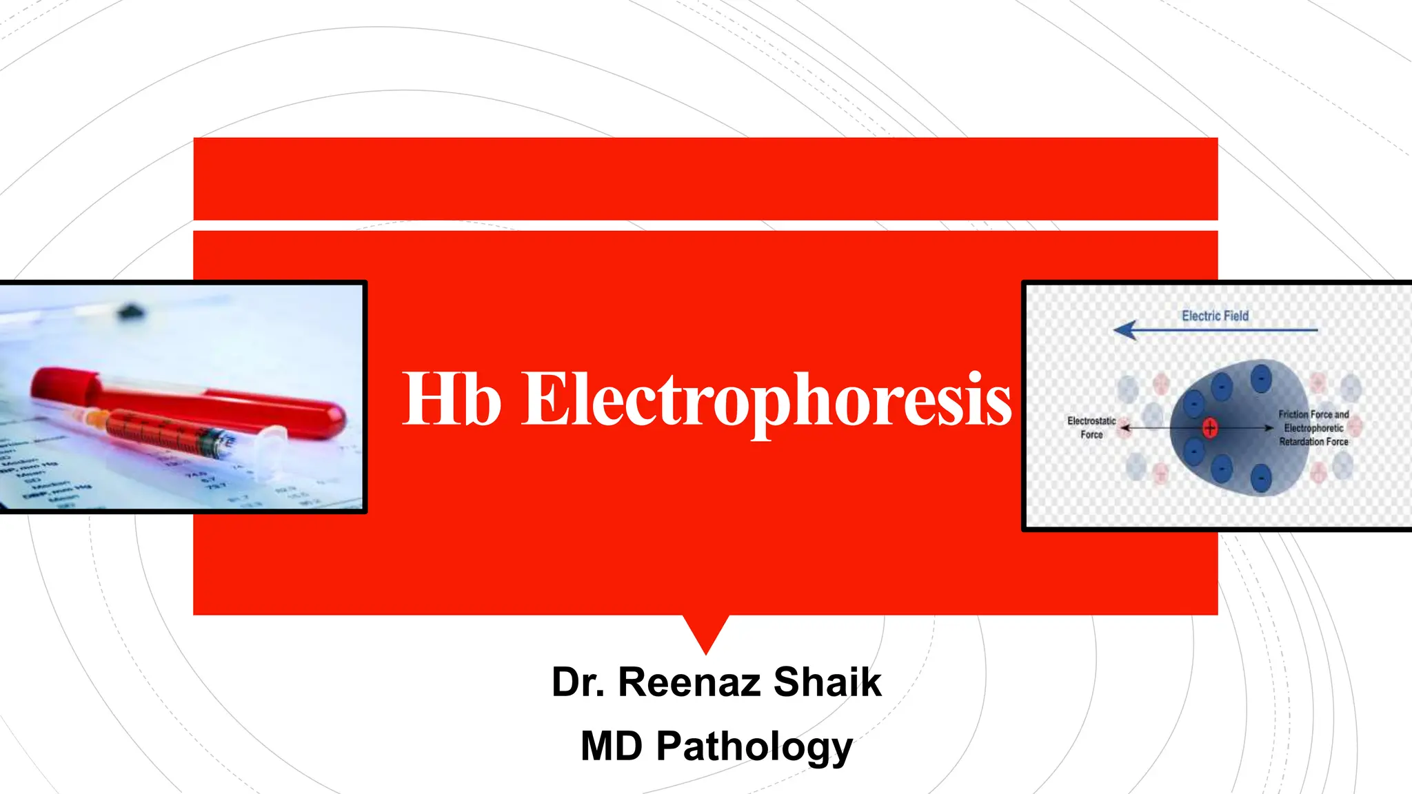
Hb electrophoresis- Types, Procedure and Analysis (20)
Lung Abscess and Pneumonia (Pathology)Reenaz Shaik
Lung abscess is formed due to necrosis within the pulmonary parenchyma resulting in the formation of cavities.
Pneumonia is due to inflammatory response in lung parenchyma distal to the terminal bronchioles.
Wound Healing is a natural physiological reaction to tissue injury. It involves numerous cell types, cytokines, mediators. Understanding basic wound healing will help in identifying molecular level target genes that can enhance and expedite natural wound healing
Westgard's rules and LJ (Levey Jennings) Charts.Reenaz Shaik
Quality Control is a process used to monitor and evaluate the analytical process that produces patients results. Planning, documenting and agreeing on a set of guidelines ensures quality.
Myeloproliferative disorders describe a group of disorders that result from unchecked, autonomous clonal proliferation of cellular elements of the bone marrow.
JMML is a rare cancer of blood that affects young children. There is a sustained abnormal and excessive production of myeloid progenitors and monocytes.
High Profile"*Call "*Girls in Kolkata ))86-075-754-83(( "*Call "*Girls in Kol...Nisha Malik Chaudhary
High Profile "*Call "*Girls in Kolkata ))86-075-754-83(( "*Call "*Girls in Kolkata Available
Kolkata "Call "Girls 74046-34175 "Call "Girl Number in Kolkata | A nutshell review for Hot "Call "Girls in Kolkata . MY experience was superb with them this is the only recommended "Call "Girls service in Kolkata "Call "Girls and again then Russian. so overall my practice was magnificent. The price is also moderate per hour. The plus point is the "Girl comes instantly to your lo"Cation doesn't matter you are in Bur Kolkata or al Nahda or Kolkata or any area she comes undeviatingly to your hotel room. Definitely recommend the "Call "Girls agency. A nutshell review for Hot "Call "Girls in Kolkata . MY experience was superb with them this is the only recommended "Call "Girls service in Kolkata with verified "Call "Girls . I am using their services from past 6 months they never ever disappointed me in any way. Let's just say if i asked them to provide me russian "Call "Girls they fulfilled my request or even beautiful "Call "Girls or indian "Call "Girls in Kolkata . They have their owen drivers who brings the "Call "Girls in less time in any area of Kolkata like bur Kolkata marina or jumeirah or even in jebel ali as well. I'm writing here everything after experience their services in all conditions.
Mainstreaming #CleanLanguage in healthcare.pptxJudy Rees
In healthcare, every day, millions of conversations fail. They fail to cover what’s really important, fail to resolve key issues, miss the point and lead to misunderstandings and disagreements.
Clean Language is one approach that can improve things. It’s a set of precise questions – and a way of asking them – which help us all get clear on what matters, what we’d like to have happen, and what’s needed.
Around 1000 people working in healthcare have trained in Clean Language skills over the past 20+ years. People are using what they’ve learnt, in their own spheres, and share anecdotes of significant successes. But the various local initiatives have not scaled, nor connected with each other, and learning has not been widely shared.
This project, which emerged from work done by the NHS England South-West End-Of-Life Network, with help from the Q Community and especially Hesham Abdalla, aims to fix that.
Chemical kinetics is the study of the rates at which chemical reactions occur and the factors that influence these rates.
Importance in Pharmaceuticals: Understanding chemical kinetics is essential for predicting the shelf life of drugs, optimizing storage conditions, and ensuring consistent drug performance.
Rate of Reaction: The speed at which reactants are converted to products.
Factors Influencing Reaction Rates:
Concentration of Reactants: Higher concentrations generally increase the rate of reaction.
Temperature: Increasing temperature typically increases reaction rates.
Catalysts: Substances that increase the reaction rate without being consumed in the process.
Physical State of Reactants: The surface area and physical state (solid, liquid, gas) of reactants can affect the reaction rate.
Hepatocarcinoma today between guidelines and medical therapy. The role of sur...Gian Luca Grazi
Today more than ever, hepatocellular carcinoma therapy is experiencing profound and substantial changes.
The association atezolizumab (ATEZO) plus bevacizumab (BEVA) has demonstrated its effectiveness in the post-operative treatment of patients, improving the results that can be achieved with liver resections. This after the failure of the use of sorafenib in the already historic STORM study.
On the other hand, the prognostic classification of BCLC is now widely questioned. It is now well recognized that the indications for surgery for patients with hepatocellular carcinoma are certainly narrow in BCLC and no longer reflect what is common everyday clinical practice.
Today, the concept of multiparametric therapeutic hierarchy, which makes the management of patients with hepatocellular carcinoma much more flexible and allows the best therapy for the individual patient to be identified based on their clinical characteristics, is gaining more and more importance.
The presentation traces these profound changes that are taking place in recent years and offers a modern vision of the management of patients with hepatocellular carcinoma.
Coronary Circulation and Ischemic Heart Disease_AntiCopy.pdfMedicoseAcademics
In this lecture, we delve into the intricate anatomy and physiology of the coronary blood supply, a crucial aspect of cardiac function. We begin by examining the physiological anatomy of the coronary arteries, which lie on the heart's surface and penetrate the cardiac muscle mass to supply essential nutrients. Notably, only the innermost layer of the endocardial surface receives direct nourishment from the blood within the cardiac chambers.
We then explore the specifics of coronary circulation, including the dynamics of blood flow at rest and during strenuous activity. The impact of cardiac muscle compression on coronary blood flow, particularly during systole and diastole, is discussed, highlighting why this phenomenon is more pronounced in the left ventricle than the right.
Regulation of coronary circulation is a complex process influenced by autonomic and local metabolic factors. We discuss the roles of sympathetic and parasympathetic nerves, emphasizing the dominance of local metabolic factors such as hypoxia and adenosine in coronary vasodilation. Concepts like autoregulation, active hyperemia, and reactive hyperemia are explained to illustrate how the heart adjusts blood flow to meet varying oxygen demands.
Ischemic heart disease is a major focus, with an exploration of acute coronary artery occlusion, myocardial infarction, and subsequent physiological changes. The lecture covers the progression from acute occlusion to infarction, the body's compensatory mechanisms, and the potential complications leading to death, such as cardiac failure, pulmonary edema, fibrillation, and cardiac rupture.
We also examine coronary steal syndrome, a condition where increased cardiac activity diverts blood flow away from ischemic areas, exacerbating the condition. The long-term impact of myocardial infarction on cardiac reserve is discussed, showing how the heart's capacity to handle increased workloads is significantly reduced.
Angina pectoris, a common manifestation of ischemic heart disease, is analyzed in terms of its causes, presentation, and referred pain patterns. We identify factors that exacerbate anginal pain and discuss both medical and surgical treatment options.
Finally, the lecture includes a case study to apply theoretical knowledge to a practical scenario, helping students understand the real-world implications of coronary circulation and ischemic heart disease. The role of biochemical factors in cardiac pain and the interpretation of ECG changes in myocardial infarction are also covered.
Join the leading All Range PCD Pharma Franchise in India and grow your business with a trusted partner. We offer an extensive range of high-quality pharmaceutical products, competitive pricing, and comprehensive marketing support. Benefit from our expertise, wide distribution network, and excellent customer service. Elevate your pharma business with See Ever Healthcare's proven PCD franchise model.
https://www.seeeverhealthcare.com/all-range-pcd-pharma-franchise-in-india/
Ontotext’s Clinical Trials Eligibility Design Assistant helps with one of the most challenging tasks in study design: selecting the proper patient population.
Hemodialysis: Chapter 8, Complications During Hemodialysis, Part 3 - Dr.GawadNephroTube - Dr.Gawad
- Video recording of this lecture in English language: https://youtu.be/pCU7Plqbo-E
- Video recording of this lecture in Arabic language: https://youtu.be/kbDs1uaeyyo
- Link to download the book free: https://nephrotube.blogspot.com/p/nephrotube-nephrology-books.html
- Link to NephroTube website: www.NephroTube.com
- Link to NephroTube social media accounts: https://nephrotube.blogspot.com/p/join-nephrotube-on-social-media.html
Chair, Benjamin M. Greenberg, MD, MHS, discusses neuromyelitis optica spectrum disorder in this CME activity titled “Mastering Diagnosis and Navigating the Sea of Targeted Treatments in NMOSD: Practical Guidance on Optimizing Patient Care.” For the full presentation, downloadable Practice Aids, and complete CME information, and to apply for credit, please visit us at https://bit.ly/4av12w4. CME credit will be available until June 27, 2025.
Case presentation of a 14-year-old female presenting as unilateral breast enlargement and found to have a giant breast lipoma. The tumour was successfully excised with the result that the presumed unilateral breast enlargement reverting back to normal. A review of management including a photo of the removed Giant Lipoma is presented.
POTENTIAL TARGET DISEASES FOR GENE THERAPY SOURAV.pptxsouravpaul769171
Theoretically, gene therapy is the permanent solution for genetic diseases. But it has several complexities. At its current stage, it is not accessible to most people due to its huge cost. A breakthrough may come anytime and a day may come when almost every disease will have a gene therapy Gene therapy have the potential to revolutionize the practice of medicine.
Hemodialysis: Chapter 8, Complications During Hemodialysis, Part 2 - Dr.GawadNephroTube - Dr.Gawad
- Video recording of this lecture in English language: https://youtu.be/FHV_jNJUt3Y
- Video recording of this lecture in Arabic language: https://youtu.be/D5kYfTMFA8E
- Link to download the book free: https://nephrotube.blogspot.com/p/nephrotube-nephrology-books.html
- Link to NephroTube website: www.NephroTube.com
- Link to NephroTube social media accounts: https://nephrotube.blogspot.com/p/join-nephrotube-on-social-media.html
2. CONTENTS
Definition and types of electrophoresis
Difference between zone and moving boundary
electrophoresis
Hb Electrophoresis: Reagents, Materials,
Methods and Procedure
Applications
Normal and Abnormal Haemoglobins
Understanding the bands
Approach
References
CONTENTS
3. Electrophoresis is the separation of charged
compounds based on their electrical charge.
An electrophoretic system consists of two electrodes
of opposite charge (anode, cathode), connected by a
conducting medium called an electrolyte.
The separation effect on the ionic particles results
from differences in their velocity (v), which is the
product of the particle's mobility (m) and the field
strength (E).
DEFINITION
V=mE
4. USES
Biological molecules such as
Amino acids
Peptides
Proteins
Nucleotides
Nucleic acid
Inorganic material can be separated
using this technique.
5. Classification of electrophoresis
1. ZONE ELECTROPHORESIS:
Paper electrophoresis
Gel electrophoresis
Thin layer electrophoresis
Cellulose acetate electrophoresis
2. MOVING BOUNDARY ELECTROPHORESIS:
Capillary electrophoresis
Isotachophoresis
Isoelectric Focussing
Immune electrophoresis
TYPES
6. DIFFERENC
ES
Zone electrophoresis: Migration of
charged molecules in a solution with the
supporting medium. The separated
components are formed like discrete zones
in the supporting medium.
Moving boundary electrophoresis:
Migration of charged molecules in a free
moving solution, without the presence of a
supporting medium.
7. HB
ELECTROPHOR
ESIS
Screening test to identify different variant
and abnormal haemoglobins.
It helps in diagnosis of diseases caused by
abnormal haemoglobins like thalassemia,
sickle cell anemia, polycythemia rubra
vera.
Helps in monitoring treatment and screen
for genetic disorders.
8. PRINCIPLE
It uses the principle of Gel electrophoresis.
Different haemoglobin have different
charges and according to those charges
and the amount of haemoglobin, different
chains move at different speed in gel are
separated.
9. HEMOGLOBIN STRUCTURE
Electrophoresis
buffer(Tris/EDTA/borate(TEB), pH 8.5)
Wetting agents (e.g., Zip zone Prep
solution)
Fixative stain/solution(Ponceau S 5 g)
Haemolysing reagents(0.5% Triton in 100
mg potassium cyanide)
De-staining solution (3% acetic acid)
REAGENT
10. HEMOGLOBIN STRUCTURE
Specimen : Blood
Packed red cells are preferred ; if whole
blood used paraprotein or high concentration
of polyclonal Ig may produce a band.
MATERIAL
11. HEMOGLOBIN STRUCTURE
Cellulose acetate (CA) electrophoresis
Alkaline electrophoresis
Citrate Agar electrophoresis
Alkaline and Citrate Agar electrophoresis
are commonly used methods
METHODS
12. STEPS
FIRST STEP: Cellulose acetate at alkaline
pH (8.4) done as an initial procedure. •
Separation is largely determined by electrical
charge. • At this pH, Hb is negatively
charged and moves toward the positively
charged anode.
SECOND STEP: Citrate agar or agarose gel
at acid pH (6)
13. HEMOGLOBIN STRUCTURE
Sample Collection(blood)
Centrifuge sample at 1200 g for 5 mins.
Prepare the electrophoresis tank with TEB
buffer
Soak the cellulose acetate into buffer for 5
mins
Fill the sample well plate with 5 μl of each
diluted sample and cover with glass slide to
prevent evaporation
Load a second sample well plate with Zip
Zone Prep solution
PROCEDURE
14. HEMOGLOBIN STRUCTURE
Then Apply them to a blotter
Blot the cellulose acetate strip twice
between two layers of clean blotting paper
Do not allow the cellulose acetate to dry
Load the applicator by depressing the tips
into the sample wells twice
Place the cellulose acetate plates across
the bridges
After 25 mins of electrophoresis
immediately transfer the cellulose acetate
to ponceau S and fix and stain for 5 mins
PROCEDURE
Cont..
15. HEMOGLOBIN STRUCTURE
Remove excess stain by washing for 5 min in the
first acetic acid reservoir
Label the membrane and store in protective
envelope
PROCEDURE
Cont..
18. HEMOGLOBIN STRUCTURE
Hematoma
Bleeding
Infection at the puncture site
Fainting or feeling lightheaded
Swelling(also called phlebitis)
RISKS
21. ABNORMAL
Hb’s RESULT
Abnormal Hb Manifesting Disease
High Hb A2 and Hb F Mild form of Thalassemia
High Hb F Hereditary Persistence of Foetal
Haemoglobin
High Hb S Sickle Cell Disease
High Hb C Anemia and enlarged spleen
High Hb E Anemia and small RBC’s
Hb DD, Hb SD Sickle disorder
Hb CC, Hb SC, Hb SS Mild, Moderate, Severe SCD
Hb H • Moderate to severe anemia,
jaundice, splenomegaly
• Blood: microcytosis,
hypochromia, target cells,
polychromasia
Hb Lepore- Boston Mild Thalassemia minor
Hb Koln Mild congenital hemolytic
anemia (AD, maybe mistaken for
hereditary spherocytosis) •
Hypochromia, macrocytosis •
Broad smudge in the S position
22. HEMOGLOBIN STRUCTURE
Reasons you may not be able to have the
test or why the results may not be helpful
include:
Having a blood transfusion in the last 3
months
Having Iron deficiency anemia. This can
cause falsely low results for
haemoglobin A2
WHAT
AFFECTS
THE TEST?
23. HEMOGLOBIN STRUCTURE
Evaluation of unexplained haemolytic
anemia
Microcytic anemia unrelated to iron
deficiency, chronic disease, or lead toxicity
A peripheral smear with abnormal red cell
features
Positive family history of
haemoglobinopathy
Positive neonatal screen results
Positive results on sickle cell or solubility
test
APPLICATIONS
24. There are different forms of normal
(e.g. HbA, Hb F, Hb A2)
Abnormal (e.g. Hb S, Hb C, Hb D, Hb
G) hemoglobins.
HAEMOGLOBINS
30. HEMOGLOBINOPATH
YAPROACH
This list shows some of the commoner tests
used to investigate the hemoglobinopathies.
Blood count
Haemoglobin electrophoresis: Cellulose
acetate pH 8.4, Citrate agar pH 6
Solubility tests
Quantitation: Hb A2, Hb F, Hb Barts
Tests for unstable hemoglobins
Gene analysis