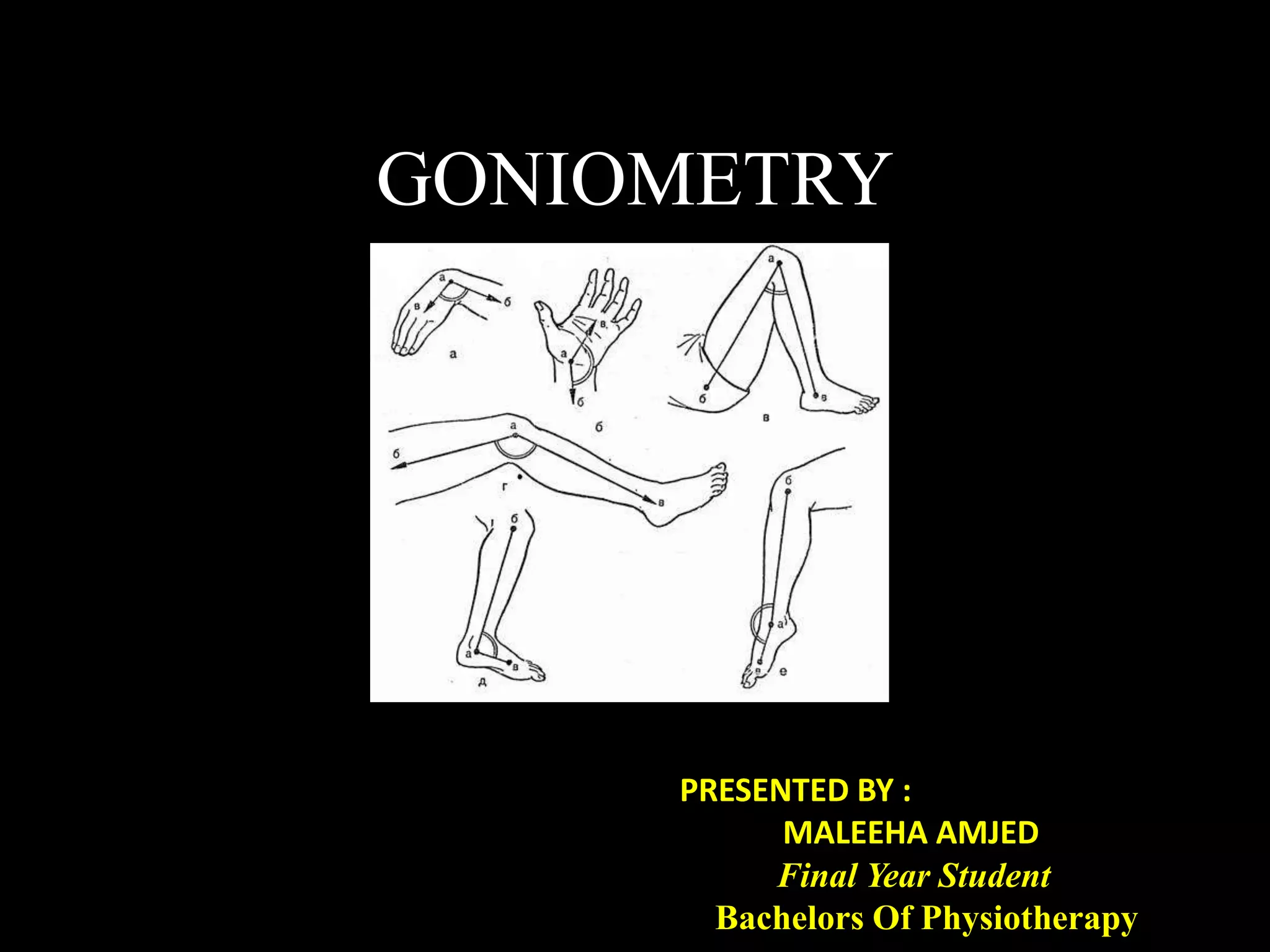
Goniometry refers to the measurement of joint angles in the human body. It is an important part of a physical examination to determine range of motion, evaluate progress, and modify treatment. There are different types of goniometers used to measure motion in various planes at joints like the shoulder, elbow, wrist, fingers, hip, and spine. Factors like a person's age, joint health, surrounding soft tissues, and pathological conditions can impact the normal range of motion values. Proper positioning, stabilization, and identification of bony landmarks is required to accurately measure and document a joint's range of motion.
1. The document discusses posture analysis and identifies key aspects to evaluate, including the spinal curves, pelvis, shoulders, and lower extremities from the lateral, posterior, and anterior views.
2. Correct posture maintains the natural curves of the spine with minimal joint stress, while poor posture can result from positional habits, muscle imbalances, or underlying medical conditions and lead to increased joint stress.
3. A thorough posture analysis examines the body with reference to plumb lines and assesses for common postural faults in each region, such as rounded shoulders, anterior pelvic tilt, or foot pronation.
Frenkel's Exercise (Bangladesh Health Profession Institute) CRPBipul Debnath
This document provides an overview and instructions for Frenkel's Exercises, which were developed in 1889 by Swiss physician Heinrich Sebastian Frenkel to treat patients with sensory ataxia and loss of proprioception. The exercises are a series of gradual, progressive movements designed to improve coordination, control, and confidence in movement. The document describes the indications, principles, techniques, and specific exercises for the lower and upper extremities in different positions like lying, sitting, and standing. It also discusses factors that affect the exercises and how to progress the routine based on the patient's disability level and control.
Rood's approach is a neurophysiological technique developed in 1940 based on reflex models of motor control. It uses sensory stimulation to normalize tone and elicit desired muscle responses based on developmental sequences. The key concepts are:
1. Categorizing muscles as tonic or phasic for stability or mobility.
2. Using ontogenic sequences of motor and vital functions development.
3. Applying appropriate sensory stimuli like touch or vibration to proprioceptive, exteroceptive, and vestibular receptors.
4. Manipulating the autonomic nervous system with techniques like icing or warming.
This document discusses end feel and range of motion measurements. It defines end feel as the quality of movement perceived by the practitioner at the end of available range of motion, which can provide information about joint structures. End feel is classified as soft, firm, or hard. The document then provides examples of normal end feel for various joints and describes techniques for measuring range of motion for major joints, including goniometer positioning and patient positioning.
This document describes various mat activities (MAT) used in physical therapy. It discusses 9 principles of MAT including concentration, control, fluidity, etc. It then describes different MAT positions and exercises including rolling, prone on elbows, prone on hands, supine on elbows, pull ups, lifting, quadruped position, kneeling, and sitting. The goals of MAT are to facilitate balance, promote stability, mobilize and strengthen the trunk and limbs, and train for functional activities. Details are provided on how to perform several example MAT exercises and positions.
This document discusses balance, fall prevention, and balance assessment and training. It defines balance as control of the center of mass over the base of support. Age-related changes and diseases that impact balance components are reviewed. Valid tools to measure balance include the Berg Balance Scale, Timed Up and Go test, and Functional Reach test. Balance training exercises discussed include calf stretches, heel/toe raises, soft surface stance, and exercises using movable surfaces like Swiss balls and tilt boards. Both hard and soft surfaces are used to challenge static and dynamic balance.
Spasticity, rigidity, hypotonia, dystonia, decerebrate rigidity, and decorticate rigidity are abnormal tones that can occur. Examination of tone includes initial observation, passive and active motion testing using scales like the Modified Ashworth Scale. Typical patterns of spasticity in upper and lower limbs are described for upper motor neuron lesions.
This document discusses the kinetics and kinematics of human gait. It defines kinetics as the study of forces acting on bodies, and kinematics as the study of motion without regard to forces. The document outlines the major forces involved in gait including externally generated forces like gravity and ground reaction forces, and internally generated forces from muscle contraction. It describes the motions and forces at the ankle, knee, and hip joints throughout the gait cycle. Measurement techniques for kinetics like force plates and for kinematics like motion capture are also summarized.
Myofascial release refers to the manual
technique for stretching the fascia and
releasing bonds between fascia and
Lintegument, musles,and bones, with the goal of
eliminating pain, increasing range of motion
and balancing the body.
The document discusses gait and the gait cycle. It defines gait as a person's pattern of walking and notes walking patterns can differ between individuals. The gait cycle is defined as the period from one heel strike to the next heel strike of the same limb. The gait cycle consists of the stance phase, when the foot is on the ground, and the swing phase, when the foot is off the ground. Temporal and distance variables are used to analyze gait, including single limb support time, stride length, and degree of toe out. The document also reviews the kinematics and kinetics of normal gait.
The document discusses various mobility devices used to increase patient mobility. It describes devices from ancient times like canes made from tree branches to modern devices with aluminum, steel, plastic and rubber. Key devices discussed include canes, crutches, walkers, wheelchairs and scooting boards. The document provides details on appropriate usage, measurements, adjustments and positioning for different mobility devices.
Stretching involves applying tension to muscles and connective tissues to increase flexibility and range of motion. There are several types of stretching including static, cyclic, ballistic, PNF, and mechanical. The key factors in stretching are proper alignment, stabilization, low intensity, and long duration to minimize muscle resistance and maximize tissue elongation. Stretching can be done manually, through self-stretching exercises, or using mechanical devices.
This document discusses joint mobility and range of motion exercises. It defines types of range of motion including active, passive, and active-assisted. It describes causes of limited mobility like injury, immobilization, or lifestyle. The principles, preparation, and techniques for range of motion exercises are outlined, including positioning, monitoring the patient's response, and moving joints smoothly through their pain-free range. Guidelines are provided for applying range of motion exercises to individual joints. The goals are to maintain joint mobility and function while avoiding further injury.
The document discusses principles of joint mobilization including using lower grades to reduce pain and higher grades to increase mobility. It outlines convex-concave rules for determining glide direction in different joints. Treatment glides are described to improve range of motion in various joints like the shoulder, knee, ankle and elbow. Open-packed positions and grades of movement are also defined. The goal of a joint mobilization treatment is to increase range of motion through appropriate gliding techniques.
- Stand behind patient
- Hold guarding belt posteriorly
- Provide support to trunk
Patient:
- Hold handrail with unaffected hand
- Place crutch/cane on step above
- Bring affected LE forward and place foot on step
- Bring unaffected LE forward and place foot on same step
- Repeat for each step
Descending stairs :-
• Therapist – postero-lateral on unaffected side
• Maintain wide BOS
• Take step only when patient is not moving
Patient:
- Hold handrail with unaffected hand
- Place crutch/cane on step below
- Bring unaffected LE back and place foot on step below
- Bring affected LE back and
As a general term, traction means pulling on part of the body.
Most often, traction uses mechanical force (sometimes generated by weights and pulleys) to put tension on a displaced bone or joint, such as a dislocated shoulder, to put it back in position and keep it still. In the medical field, traction refers to the practice of slowly and gently pulling on a fractured or dislocated body part. It’s often done using ropes, pulleys, and weights. These tools help apply force to the tissues surrounding the damaged area.
The document discusses the key components and phases of normal human gait. It defines gait as rhythmic alternating movements that propel the body's center of gravity forward. The gait cycle consists of stance and swing phases for each foot. Stance is 60% of the cycle from heel contact to toe off, while swing is 40% between toe off and next heel contact. Gait involves coordinated motion of the hips, knees, ankles, and toes through flexion, extension, and rotation. The center of gravity follows an arched path minimized through determinants like pelvic tilt and rotation, knee flexion, and ankle and foot interactions.
The document discusses the biomechanics of the spine. It describes the structure of the spine including the 33 vertebrae and intervertebral disks. It discusses the articulations between vertebrae including the intervertebral joints between vertebral bodies and disks, and the zygapophyseal joints between articular processes. It also describes the ligaments that connect vertebrae like the anterior and posterior longitudinal ligaments. The spine functions to provide support, stability, and mobility and withstands various forces like axial compression, tension, bending, torsion and shear stresses.
Orthosis are devices used to support weak joints and correct deformities. They work by applying three point pressure and distributing weight across a wide surface area. Common orthosis include ankle foot orthosis (AFO) which support the ankle and foot, knee ankle foot orthosis (KAFO) which stabilize the knee and lower leg, and hip knee ankle foot orthosis (HKAFO) which provide support from the hip to the foot. Orthosis are made of plastic or metal and their design depends on the joints needing support and the individual's condition.
Goniometry refers to the precise measurement of joint angles using instruments such as a universal goniometer. Goniometric data is used to determine impairment, establish diagnoses, develop treatment plans, and evaluate progress. Joint motion includes arthrokinematics (gliding and spinning of joint surfaces) and osteokinematics (bone movements). Range of motion is measured in three planes (sagittal, frontal, transverse) using instruments properly aligned with bony landmarks. Both active and passive range of motion are measured to evaluate joint integrity and flexibility. Restricted or increased range of motion can indicate conditions like capsular patterns of hypomobility or generalized hypermobility.
Range of motion (ROM) measurements are performed to evaluate joint impairment, develop treatment goals, assess progress, and modify treatment. ROM is described in 3 planes and axes and measured using a goniometer. Active ROM is voluntary motion while passive ROM uses external assistance. Several factors determine ROM including joint integrity, scarring, age, gender, joint shape, and health of surrounding tissues. Common causes of limited ROM include contractures, arthritis, and pain. Precise positioning and stabilization are needed to reliably measure ROM of various joints like the shoulder, spine, and knee. Standardized testing procedures and documentation of measurements are important.
This document provides information about goniometry and range of motion measurements of various joints, including the shoulder complex. It defines goniometry as the measurement of joint angles using a goniometer. The document describes how to position and stabilize the individual and properly align the goniometer to measure flexion and extension of the shoulder joint. Flexion and extension occur in the sagittal plane around the medial-lateral axis. Normal range of motion for shoulder flexion is 165-180 degrees and for glenohumeral flexion is 100-115 degrees.
Goniometry is used to measure joint range of motion. There are different types of goniometers including universal, finger, and electro goniometers. To take an accurate measurement, the therapist positions the goniometer arms parallel to the longitudinal axis of the proximal and distal body parts, with the axis over the joint. Range of motion can then be measured actively or passively. Goniometry is used to assess limitations, track progress, and guide treatment for conditions affecting joint mobility.
This document provides an overview of upper extremity orthoses, including their objectives, nomenclature, designs, and specific examples. The main objectives of upper limb orthoses are protection, correction, and assistance. They are named based on the joints they cover, their function, condition treated, appearance, or designer. Designs include non-articular, static, serial static, dynamic, and more. Examples provided include wrist splints, elbow braces, shoulder slings, and finger orthoses. The document aims to classify and describe the various types of upper extremity splints and braces used in orthotic treatment.
Goniometry refers to the measurement of joint angles using a goniometer. There are various types of goniometers that have a body and two arms to align along bones proximal and distal to the joint. Goniometry is used to measure both active and passive range of motion of joints to assess limitations. The document provides details on goniometry procedures, principles, factors affecting range of motion, indications, contraindications and examples of normal range of motion measurements for various upper and lower limb joints.
This document defines goniometry as a technique used to measure range of motion in joints. It discusses the definition, uses, parts of a goniometer, degrees of freedom in joints, and procedures for goniometric measurement. Key points covered include that a goniometer consists of stationary, moving, and body arms to measure angles in degrees, and it is used to identify contractures or decreased range of motion from injury or disuse, help develop treatment goals, and evaluate rehabilitation progress. Normal ranges of motion are provided for the shoulder and elbow.
Goniometry involves measuring the range of motion of joints using a goniometer. Key steps in the goniometry procedure include positioning the joint at zero position, moving it to the end of its range of motion, palpating bony landmarks, aligning the goniometer, and recording the measurement. Valid and reliable goniometric measurements require proper stabilization of proximal joint components and identification of the end feel, or resistance felt at the end of range. The document then provides details on positioning, procedures, and normal ranges for measuring several upper extremity joints including the shoulder, elbow, forearm, wrist, and finger joints.
The document discusses ankle instability and arthrodesis. It provides details on:
1) The classification of ankle sprains as type I, II, or III based on the ligament damage. The anatomy of the ligaments stabilizing the medial and lateral sides of the ankle are described.
2) Diagnosis of ankle injuries involves physical exams like the anterior drawer test and talar tilt test as well as radiographic views. MRI may be used if pain persists.
3) Treatment includes RICE, bracing, surgery for severe or chronic cases using various reconstruction techniques depending on the ligaments injured.
4) Ankle arthrodesis is described as an option for end-stage ankle arthritis
Lateral epicondylitis, commonly known as tennis elbow, is a painful condition caused by overuse and microtears of the tendons that connect the forearm muscles to the lateral epicondyle of the humerus. The condition results in pain at the outside of the elbow. Conservative treatments include activity modification, bracing, stretching, strengthening exercises, and shock wave therapy. Surgical intervention is considered if conservative treatments fail to provide relief after 6 months.
This document provides information on various types of hand orthosis including their objectives, indications, and principles. It describes static and dynamic orthosis used to immobilize, support, correct deformities, and facilitate motion of the wrist, fingers, and thumb. Examples include cock-up splints, gauntlet immobilization splints, and dynamic wrist extension splints. Biomechanical principles like three point pressure and stress distribution are discussed. Contraindications and importance of physical therapy evaluation and training are also summarized.
The document provides guidance on assessing a patient's musculoskeletal system and rheumatological symptoms. It describes:
1. Questions to ask about joint pain, stiffness, swelling, deformities, and ability to perform activities.
2. How to perform a focused physical exam using a modified GALS (gait, arms, legs, spine) method to quickly check mobility.
3. Details on examining individual joints like the back, shoulders, hands, hips, and feet to identify areas of pain or reduced movement that could indicate arthritis.
The assessment aims to identify key symptoms, affected joints, onset and progression of issues in order to formulate potential diagnoses.
This document discusses common shoulder pathologies seen in industrial athletes. It begins with shoulder anatomy including bones and muscles. It then discusses common injuries like impingement and rotator cuff tears. Impingement is caused by encroachment in the subacromial space and can be primary from bone spurs or congenital issues, or secondary from muscle imbalances or poor posture. Rotator cuff tears can be partial or full thickness and result from repetitive stress or acute trauma. Treatment involves rehabilitation exercises and potentially surgery. Trigger points are also discussed as a potential cause of shoulder pain presenting in specific patterns that can be treated with massage or spray techniques. Overall the document provides an overview of shoulder issues in industrial settings and potential
1. The document provides information on examining the shoulder, elbow, wrist, and hand, including anatomy, inspection, palpation, range of motion tests, and special tests.
2. Common causes of pain in these areas are described, including rotator cuff injuries, arthritis, tendinitis, bursitis, and neurological issues.
3. Examination techniques for each area include inspection for deformities, swelling, atrophy; palpation for temperature, tenderness, crepitus; and range of motion and special tests like impingement signs and drop arm test for shoulders.
This document discusses orthotics and their use in rehabilitation. It begins by describing how bioengineering devices like orthotics play an important role in orthopedic and neurological rehabilitation by improving function and support. It then discusses different types of orthotics in more detail, including their components, classifications, indications for use, and general principles. Specific orthotics for the ankle, knee, and hip are also outlined.
The document discusses goniometry, which is the measurement of joint angles using a goniometer. It outlines what goniometry is, the importance and types of goniometers, how to measure range of motion for various joints including the shoulder, wrist, hip and hand, and considerations for validity and reliability when performing goniometric measurements. Proper procedures and positioning for accurate goniometric assessment of different joints are described.
The document discusses goniometry, which is the measurement of joint angles using a goniometer. It outlines what goniometry is, the importance and types of goniometers, how to measure range of motion for various joints including the shoulder, wrist, hip and hand, and issues around validity and reliability. Standard procedures for goniometric measurement are provided along with important notes for accurate assessment.
Chair and Presenter, Stephen V. Liu, MD, Benjamin Levy, MD, Jessica J. Lin, MD, and Prof. Solange Peters, MD, PhD, discuss NSCLC in this CME/MOC/NCPD/AAPA/IPCE activity titled “Decoding Biomarker Testing and Targeted Therapy in NSCLC: The Complete Guide for 2024.” For the full presentation, downloadable Practice Aids, and complete CME/MOC/NCPD/AAPA/IPCE information, and to apply for credit, please visit us at https://bit.ly/4bBb8fi. CME/MOC/NCPD/AAPA/IPCE credit will be available until July 1, 2025.
High Profile"*Call "*Girls in Kolkata ))86-075-754-83(( "*Call "*Girls in Kol...Nisha Malik Chaudhary
High Profile "*Call "*Girls in Kolkata ))86-075-754-83(( "*Call "*Girls in Kolkata Available
Kolkata "Call "Girls 74046-34175 "Call "Girl Number in Kolkata | A nutshell review for Hot "Call "Girls in Kolkata . MY experience was superb with them this is the only recommended "Call "Girls service in Kolkata "Call "Girls and again then Russian. so overall my practice was magnificent. The price is also moderate per hour. The plus point is the "Girl comes instantly to your lo"Cation doesn't matter you are in Bur Kolkata or al Nahda or Kolkata or any area she comes undeviatingly to your hotel room. Definitely recommend the "Call "Girls agency. A nutshell review for Hot "Call "Girls in Kolkata . MY experience was superb with them this is the only recommended "Call "Girls service in Kolkata with verified "Call "Girls . I am using their services from past 6 months they never ever disappointed me in any way. Let's just say if i asked them to provide me russian "Call "Girls they fulfilled my request or even beautiful "Call "Girls or indian "Call "Girls in Kolkata . They have their owen drivers who brings the "Call "Girls in less time in any area of Kolkata like bur Kolkata marina or jumeirah or even in jebel ali as well. I'm writing here everything after experience their services in all conditions.
Pharmacotherapy of Asthma and Chronic Obstructive Pulmonary Disease (COPD)HRITHIK DEY
This PowerPoint presentation provides an in-depth overview of the pharmacotherapy approaches for managing asthma and Chronic Obstructive Pulmonary Disease (COPD). It covers the pathophysiology of these respiratory conditions, the various classes of medications used, their mechanisms of action, indications, side effects, and the latest treatment guidelines. Designed for students, healthcare professionals, and anyone interested in respiratory pharmacology, this presentation offers a comprehensive understanding of current therapeutic strategies and advancements in the field.
Case presentation of a 14-year-old female presenting as unilateral breast enlargement and found to have a giant breast lipoma. The tumour was successfully excised with the result that the presumed unilateral breast enlargement reverting back to normal. A review of management including a photo of the removed Giant Lipoma is presented.
Hemodialysis: Chapter 8, Complications During Hemodialysis, Part 2 - Dr.GawadNephroTube - Dr.Gawad
- Video recording of this lecture in English language: https://youtu.be/FHV_jNJUt3Y
- Video recording of this lecture in Arabic language: https://youtu.be/D5kYfTMFA8E
- Link to download the book free: https://nephrotube.blogspot.com/p/nephrotube-nephrology-books.html
- Link to NephroTube website: www.NephroTube.com
- Link to NephroTube social media accounts: https://nephrotube.blogspot.com/p/join-nephrotube-on-social-media.html
Causes Of Tooth Loss
PERIODONTAL PROBLEMS ( PERIODONTITIS, GINIGIVITIS)
Systemic Causes Of Tooth Loss
1. Diabetes Mellitus
2. Female Sexual Hormones Condition
3. Hyperpituitarism
4. Hyperthyroidism
5. Primary Hyperparathyroidism
6. Osteoporosis
7. Hypophosphatasia
8. Hypophosphatemia
Causes Of Tooth Loss
CARIES/ TOOTH DECAY
Causes Of Tooth Loss
CAUSES OF TOOTH LOSS
Consequence of tooth loss
Anatomic
Loss of ridge volume both height and width
Bone loss :
mandible > maxilla
Posteriorly > anteriorly
Anatomic consequences
Broader mandibular arch with constricting maxilary arch
Attached gingiva is replaced with less keratinised oral mucosa which is more readily traumatized.
Anatomic consequences
Tipping of the adjacent teeth
Supraeruption of the teeth
Traumatic occlusion
Premature occlusal contact
Anatomic Consequences
Physiologic consequences
Physiologic Consequences
Decreased lip support
Decreased lower facial height
Physiologic Consequences
Physiologic consequences
Education of Patient
Diagnosis, Treatment Planning, Design, Treatment, Sequencing, and Mouth Preparation
Support for Distal Extension Denture Bases
Establishment and Verification of Occlusal Relations and Tooth Arrangements
Initial Placement Procedures
Periodic Recall
Education of Patient
Informing a patient about a health matter to
secure informed consent.
Patient education should begin at the initial
contact with the patient and should continue throughout treatment.
The dentist and the patient share responsibility for the ultimate success of a removable partial denture.
This educational procedure is especially important when the treatment plan and prognosis are discussed with the patient.
Diagnosis, Treatment Planning, Design, Treatment, Sequencing, and Mouth Preparation
Begin with thorough medical and dental histories.
The complete oral examination must include both clinical and radiographic interpretation of:
caries
the condition of existing restorations
periodontal conditions
responses of teeth (especially abutment teeth) and residual ridges to previous stress
The vitality of remaining teeth
Continued…..
Occlusal plan evaluation
Arch form
Evaluation of Occlusal relationship through mounting the diagnostic cast
The dental cast surveyor is an absolute necessity in which patients are being treated with removable partial dentures.
Mouth preparations, in the appropriate sequence, should be oriented toward the goal of
providing adequate support, stability,
retention, and
a harmonious occlusion for the partial denture.
Support for Distal Extension Denture Bases
A base made to fit the anatomic ridge form does not provide adequate support under occlusal loading.
The base may be made to fit the form of the ridge when under function.
Support for Distal Extension Denture Bases
This provides support
EXPERIMENTAL STUDY DESIGN- RANDOMIZED CONTROLLED TRIALRishank Shahi
Randomized controlled clinical trial is a prospective experimental study.
It essentially involves comparing the outcomes in two groups of patients treated with a test treatment and a control treatment, both groups are followed over the same period of time. Prepare a plan of study or protocol
a. Define clear objectives
b. State the inclusion and exclusion criteria of case
c. Determine the sample size, place and period of study
d. Design of trial (single blind, double blind and triple blind method)
2. Define study population: Most often the patients are chosen from hospital or from the community. For example, for a study for comparison of home and sanatorium treatment, open cases of tuberculosis may be chosen.
3. Selection of participants by defined criteria as per plan:
Selection of participants should be done with precision and should be precisely stated in writing so that it can be replicated by others. For example, out of open cases of tuberculosis those who fulfill criteria for inclusion may be selected (age groups, severity of disease and treatment taken or not, etc.)
Randomization ensures that participants have an equal chance to be assigned to one of two or more groups:
One group gets the most widely accepted treatment (standard treatment/ gold standard)
The other gets the new treatment being tested, which researchers hope and have reason to believe will be better than the standard treatment
Subject variation: First, there may be bias on the part of the participants, who may subjectively feel better or report improvement if they knew they were receiving a new form of treatment.
Observer bias: The investigator measuring the outcome of a therapeutic trial may be influenced if he knows beforehand the particular procedure or therapy to which the patient has been subjected.
Evaluation bias: There may be bias in evaluation - that is, the investigator(Analyzer) may subconsciously give a favorable report of the outcome of the trial.
Co-intervention:
participants use other therapy or change behavior
Study staff, medical providers, family or friends treat participants differently.
Biased outcome ascertainment:
participants may report symptoms or outcomes differently or physicians
Investigators may elicit symptoms or outcomes differently
A technique used to prevent selection bias by concealing the allocation sequence from those assigning participants to intervention groups, until the moment of assignment.
Allocation concealment prevents researchers from influencing which participants are assigned to a given intervention group.
All clinical trials must be approved by Institutional Ethics Committee before initiation
It is mandatory to register clinical trials with Clinical Trials Registry of India
Informed consent from all study participants is mandatory.
A preclinical trial is a stage of research that begins before clinical trials, and during which important feasibility and drug safety data are collected.
Following points high.
Mainstreaming #CleanLanguage in healthcare.pptxJudy Rees
In healthcare, every day, millions of conversations fail. They fail to cover what’s really important, fail to resolve key issues, miss the point and lead to misunderstandings and disagreements.
Clean Language is one approach that can improve things. It’s a set of precise questions – and a way of asking them – which help us all get clear on what matters, what we’d like to have happen, and what’s needed.
Around 1000 people working in healthcare have trained in Clean Language skills over the past 20+ years. People are using what they’ve learnt, in their own spheres, and share anecdotes of significant successes. But the various local initiatives have not scaled, nor connected with each other, and learning has not been widely shared.
This project, which emerged from work done by the NHS England South-West End-Of-Life Network, with help from the Q Community and especially Hesham Abdalla, aims to fix that.
JMML is a rare cancer of blood that affects young children. There is a sustained abnormal and excessive production of myeloid progenitors and monocytes.
3. What is Goniometry?
• The term goniometry is derived from two Greek words :
Gonia-metron
• Therefore, goniometry refers to the measurement of angles, in
particular the measurement of angles created at human joints
by the bones.
ANGLE MEASURE
4. PARTS OF MOTOR EXAMINATION
1. Nutrition Of Muscle
2. Muscle Tone
3. Reflexes
4. Range Of Motion and TCD’s
5. Manual Muscle Testing
5. Why Is It Performed ?
• Determining the presence of joint impairment
• Developing treatment goals.
• Evaluating progress or lack of progress.
• Modifying treatment.
• Motivating the subject.
• Research
7. PLANES AND AXIS
• Osteo-kinematic motions are described to be taking place in 3
cardinal planes and axis
9. Synovial joint
• Most evolved & hence most mobile type of
joints
• The ends of bony components are free to
move in relation to one another
• Bony components are indirectly connected to
one another by means of a joint capsule that
encloses the joint
10. Joint Ranges
Active ROM Passive ROM
• Active motion is the unassisted voluntary
movement of a joint. (Quality of ROM)
• Passive motion is attained by the examiner
without the patient’s assistance. (Quantity of ROM
)
• ** Normally, PROM is slightly greater than AROM
because joints have a small amount of motion at
the end range that is not under voluntary control.
12. physiologic motion is limited
by a physiologic barrier
tension develops within the
surrounding tissues
(joint capsule, ligaments
and connective tissue)
13. additional amount of passive
range of motion can be performed the anatomic
barrier cannot be
exceeded without
disrupting the
joints integrity
16. ACTIVE INSUFFICIENCY?
Flex your wrist
completely
Attempt to
tighten your
fist
• A muscle
cantcontractmaximally
across both joints
together
Much force
than in
slightly
extended
position
• The multi joint long
finger flexors enter
active insufficiency
when wrist flexes
• Shortest possible
length of muscle
17. PASSIVE INSUFFICIENCY?
FLEXION OF THE FINGERS
IS A RESULT OF
INSUFFICIENT
EXTENSIBILTY OF THE
FINGER FLEXORS
STRETCHED OVER
EXTENDED WRIST
EXTENSION OF THE
FINGERS IS A RESULT OF
INSUFFICIENT LENGTH OF
THE FINGER EXTENSORS
STRETCHED OVER FLEXED
WRIST
• LONGEST POSSIBLE LENGTH OF MUSCLE
• Muscle cant stretch maximally at both joints together
18. Other Examples of AI PI In Body and its
clinical relevance with Goniometry
• BICEPS : At the top of curl, (when biceps begin to smash against
forearm), when elbows are lifted
**Shortens biceps over both the shoulder & elbow blade
• Simultaneously lengthening the TRICEPS
• HAMS : When reaching to touch toes
**Lengthening felt as a stretch
• RECTUS FEMORIS : Hip flexion with knee extension(70 degree) is
less than hip flexion with knees bent (120 degree)
• GASTROCNEMIUS : Seated calf / heel raise places the
gastrocnemius into active insufficiency since the knee flexes too
much & ankle performs plantarflexion
19. MEASURING
JOINT RANGE OF MOTION
• Range Of Motion (ROM) is the arc of motion that
occurs at a joint or a series of joints.
• Three notation systems have been used to
define ROM :
1. The 0 to 180 degree system
2. The 180 to 0 degree system
3. The 360 degree system
Most commonly used is the 0 to 180 degree
notation system
20. Prerequisite Knowledge For Measuring ROM
a) Normal ROM’s (Range)
b) Joint Structure And Function
c) Recommended positioning for self and patient
d) Bony landmarks related to each joint
e) Alignment of Goniometer
f) Normal end-feel
g) Factors that can alter normal ROM
21. FACTORS DETERMINING AMOUNT OF ROM
Integrity Of
Joint
SurfaceRELIABILITY
Amount Of
Scarring
Present
AGE
GENDER
Shape Of
Articulating
Surface
Health
Of
Joint
Various
diseases/
pathological
conditions
Health Of
Surrounding
Tissues
Mobilty &
Pliabilty Of Soft
Tissue
22. Common pathological causes of ROM
Restriction
• Skin/soft tissue contracture
• Arthritis
• Fracture
• Burns
• Muscle weakness/paralysis
• Pain
• Edema
• Spasticity
• Presence of foreign body in the joint
23. Prerequisite Skills For Measuring ROM
• The therapist should be skilled in
Correct positioning (Pt/ Pt Jt/ PT And GM)
Stabilization for measurement
Palpation
Alignment
Recording measurements accurately
Documentation
24. • Visual observation of the joint and its adjacent
area is important to look for :
a) Compensatory motions
b) Posture
c) Muscle contour
d) Skin creases
e) Facial expressions
25. Testing Procedure
PLACE THE SUBJECT IN TESTING
POSITION
STABILIZE THE PROXIMAL JOINT SEGMENT
MOVE THE DISTAL JOINT SEGMENT TO ZERO STARTING POSITION. SLOWLY MOVE
THE DISTAL JOINT SEGMENT TO THE END OF PASSIVE ROM AND DETERMINE END FEEL
MAKE VISUAL ESTIMATE OF THE ROM
RETURN THE DISTAL JOINT SEGMENT TO THE STARTING POSITION
PALPATE THE BONY ANATOMICAL LANDMARKS
ALIGN THE GONIOMETER
26. READ & RECORD THE STARTING POSITION.
REMOVE THE GONIOMETER
STABILIZE THE PROXIMAL JOINT SEGMENT
MOVE THE DISTAL SEGMENT
THROUGH FULL ROM
REPLACE & REALIGN THE GONIOMETER. PALPATE THE ANATOMICAL LAND
MARKS AGAIN IF NECESSARY
READ & RECORD THE ROM
27. Joint Mobility Scale
Hyper Mobility
(Mild, Moderate,
Severe)
Exercise, Bracing
surgery
Normal mobility Normal function
Hypo Mobility
(Mild, Moderate,
Severe)
Exercise, Mobilization,
surgery
N
28. Documentation
• Hypo Mobility : A motion that does not start with 0
degree or ends prematurely indicates joint
hypomobility
Example : if knee joint has 30 degree of hypomobility in
flexion, it would be recorded as 30 – 135 deg
• Hyper Mobility : Joint hypermobility at the beginning
of the range is noted by inclusion of a zero between the
starting & ending measurements
Example : if the elbow joint has 5 degree of
hypermobility in extension and 140 degree of flexion ,
it would be recorded as 5 – 0 – 140 deg
29. Types of Goniometer
• Full Circle Manual Universal Goniometer (360)
• Half circle manual Goniometer (180)
• Gravity Goniometer :-
• a) Double Inclinometer (used for spine goniometry)
• b) Pendulum Inclinometer
• c) Bubble Goniometer
• Electrogoniometer
• Digital Goniometer
• Tape Measurements
• Smartphone Devices
• Use of malleable wires/sheets (in cases of deformities)
33. UNIVERSAL GONIOMETER
• A universal Goniometer may be constructed of
metal or plastic and it has 3 parts :-
1. Body of Goniometer
2. Stationary arm
3. Movable arm
(placed over the Joint being measured)
(aligned parallel with the longitudinal axis of the
fixed part)
(aligned parallel with the longitudinal axis of the
movable part)
36. Precautions !!!
1. Joint irritability status
2. Presence of Pain
3. Instability
4. Recent trauma
5. Is it really important to assess accurate ROM ??
37. Functional Ranges of various joint in
various activities
Walking
Stair ascending descending
Sitting
Squatting
Cross leg sitting
Self Feeding
Back reach
Neck reach
Etc….
38. ROM Required In ADL’s
ASCENDING STAIRS REQUIRES
BETWEEN
47 - 66 DEGREE OF HIP FLEXION
DEPENDING ON STAIR
DIMENSION
DESCENDING STAIRS REQUIRES AN
AVERAGE OF
21 - 36 DEGREE OF DORSIFLEXION,
86.9 - 107 DEGREE OF KNEE FLEXION
DEPENDING ON STAIR DIMENSIONS
39. Rising from a chair requires a mean range of
knee flexion of 90.1 - 95.0 degree and
full dorsiflexion ROM depending on height
of seat
Sitting in a chair with an
average seat height requires
112 degrees of hip flexion
40. Drinking from a cup requires about
130 degree of elbow flexion
36 to 52 degrees of shoulder flexion
Reaching objects on a high shelf
require
148 degrees of shoulder flexion
41. Using a telephone requires
approx 40 degrees of wrist
extension
Approximately
50 degrees of pronation
occur while reading a newspaper
Reaching behind the head
requires about
112 degrees of abduction
of the shoulder
42. END-FEEL
• The end of each motion at each joint is limited
from further movement by particular
anatomical structures.
• The type of structure that limits a joint motion
has a characteristic feel, which may be detected
by the therapist performing the passive ROM.
• This feeling, which is experienced by the
therapist as resistance or a barrier to further
motion, is called the end-feel.
43. NORMAL END-FEEL DESCRIPTION EXAMPLE
Soft Soft Tissue Approximation Knee flexion (contact
between soft tissue of
posterior leg and posterior
thigh)
Firm Muscular stretch
Capsular stretch
Ligamentous stretch
Hip flexion with knee
straight (passive elastic
tension of hamstring
muscles)
Extension of
metacarpophalangeal joints
of fingers
Forearm supination (tension
in the palmar radioulnar
ligament of the inferior
radioulnar joint)
Hard Bone contacting bone Elbow extension (olecranon
process of the ulna and
olecranon fossa of humerus)
44. ABNORMAL END-FEEL DESCRIPTION EXAMPLES
Soft Occurs sooner or later in the
ROM than is usual or in a joint
that normally has a firm or
hard end-feel . Feels boggy.
Soft tissue edema
Synovitis
Firm Occurs sooner or later in the
ROM than is usual or in a joint
that normally has a soft or
hard end-feel.
Increased muscular tonus
Capsular , muscular ,
ligamentous, and fascial
shortening
Hard Occurs sooner or later in the
ROM than is usual or in a joint
that normally has a soft or
firm end-feel. A bony grating
or bony block is felt.
Chondromalacia
Osteoarthritis
Loose bodies in joint
Myositis ossificans
Fracture
Empty No real end-feel because pain
prevents reaching end of
ROM. No resistance is felt
except for patient’s protective
muscle splinting or muscle
spasm.
Acute joint inflammation
Bursitis
Abscess
Fracture
Psychogenic disorder
45. JOINT MOTION TESTING
POSITION
STABILIZATION MEASUREMENTS
CERVICAL • FLEXION
• EXTENSION
• SIDE FLEXION
• ROTATION
Sitting Shoulder & chest
Shoulder & chest
to prevent
extension of
thoracic &
lumbar spine
To prevent side
flexion of
thoracic &
lumbar spine
To prevent
rotation of
thoracic &
lumbar spine
1 cm– 4.3 cm
18.5 cm–22.4cm
10.7cm-12.9cm
11cm-13.2cm
TAPE MEASUREMENTS OF THE SPINE
46. JOINT MOTION TESTING
POSITION
STABILIZATION MEASUREMENTS
THORACIC • FLEXION
• EXTENSION
• LATERAL
FLEXION
• ROTATION
STANDING
•If the subject
has balance
problems or
muscle weakness
in the LE,
measurement
can be taken in
prone/side lying
SITTING
PELVIS
To prevent
anterior tilting
To prevent
posterior tilting
To prevent lateral
tilting
To prevent
rotation
10 cms (4 inches)
15.9cm for rt LF
16.9cm for lt LF
45 degree
(universal
goniometer)
47. JOINT MOTION TESTING
POSITION
STABILIZATION MEASUREMENTS
LUMBAR • FLEXION
•EXTENSION
•LATERAL
FLEXION
STANDING PELVIS
To prevent
anterior tilting
To prevent
posterior tilting
To prevent
lateral tilting
6.7cm in males
5.8cm in females
Average
6.3cm-6.9cm
(Modified
Schober test)
1.6cm (Modified
Schober Test)
25 – 30 degree
by AMA (double
inclinometer)
49. Capsular & Non-capsular Pattern Of
Movement Restriction
• Cyriax proposed that pathological conditions
involving the entire joint capsule cause a
particular pattern of restriction involving most
of the passive motions of the joint. This pattern
is called as capsular pattern
• Restriction caused by condition involving
structures other than the entire joint capsule is
called as non-capsular pattern
• Example – Adhesive Capsulitis Shoulder
64. CERVICAL SPINE
JOINT ROM
Flexion 0º to 45º
Extension 0º to 45º
Lateral flexion 0º to 45º
Rotation 0º to 60º
THORACIC AND LUMBAR
SPINE
JOINT ROM
Flexion 0º to 80º
Extension 0º to 30º
Lateral flexion 0º to 40º
Rotation 0º to 45º
65. SHOULDER
JOINT ROM
Flexion 0º to 180º
Extension 0º to 60º
Abduction 0º to 180º
Adduction 0º
Horizontal abduction 0º to 40º
Horizontal Adduction 0º to 130º
Internal rotation
Arm in Abduction 0º to 70º
Arm in Adduction 0º to 60º
External rotation
Arm in Abduction 0º to 90º
Arm in Adduction 0º to 80º
66. ELBOW
JOINT ROM
Flexion 0º to 135º - 150º
Extension 0º
FOREARM
JOINT ROM
Pronation 0º to 80º - 90º
Supination 0º to 80º - 90º
67. WRIST
JOINT ROM
Flexion 0º to 80º
Extension 0º to 70º
Ulnar
deviation
(adduction)
0º to 30º
Radial
deviation
(abduction)
0º to 20º
THUMB
JOINT ROM
DIP flexion 0º to 80º - 90º
MCP flexion 0º to 50º
Adduction, radial
and palmar
0º
Palmar
abduction
0º to 50º
Radial abduction
Opposition
0º to 50º
68. FINGERS
JOINT ROM
MCP flexion 0º to 90º
MCP hyperextension 0º to 15º - 45º
PIP flexion 0º to 110º
DIP flexion 0º to 80º
abduction 0º to 25º
69. HIP
JOINT ROM
Flexion 0º to 120º (bent
knee)
Extension 0º to 30º
Abduction 0º to 40º
Adduction 0º to 35º
Internal rotation 0º to 45º
External rotation 0º to 45º
KNEE
JOINT ROM
Flexion 0º to 135º
70. ANKLE AND FOOT
JOINT ROM
Plantar flexion 0º to 50º
Dorsiflexion 0º to 15º
Inversion 0º to 35º
Eversion 0º to 20º
71. SOURCES
• Measurement of Joint Motion : A Guide
to Goniometry, 4th Edition, by Cynthia C. Norkin
• Physical Rehabilitation 6th Edition SuSan B.
O’Sullivan
• Magee (2002). Orthopedic physical Assessment (4th
ed.). Phil: Saunders.
• Kisner C, & Colby LA (2002). Therapeutic
exercise: Foundations and techniques (4th ed.). PA:
FA Davis.
• The Principles of Exercise Therapy (Fourth
Edition): M. Dena Gardiner.